International Journal of Medical Sciences
ISSN: 1449-1907
3.2
Impact Factor
ISSN: 1449-1907
- Current Issue
- Volume 23; 2026
- Volume 22; 2025
- Volume 21; 2024
- Volume 20; 2023
- Volume 19; 2022
- Archive
- Editorial Board
- Cover Images
- Index & Coverage
- Cover Suggestion
- Special Issues
Top
1. INTRODUCTION
2. AIM
3. OSTEOLOGY
4. TAPHONOMY
5. TAPHONOMIC IRONY: WHY...
6. THE REST OF THE FAMILY:...
7. THE CASE FOR ARRHIDAIOS AND...
8. SUMMARY AND CONCLUSION
9. PERSPECTIVES
10. ACKNOWLEDGEMENTS
References
Footnotes
1. INTRODUCTION
2. AIM
3. OSTEOLOGY
4. TAPHONOMY
5. TAPHONOMIC IRONY: WHY...
6. THE REST OF THE FAMILY:...
7. THE CASE FOR ARRHIDAIOS AND...
8. SUMMARY AND CONCLUSION
9. PERSPECTIVES
10. ACKNOWLEDGEMENTS
References
Footnotes

 Global reach, higher impact
Int J Med Sci 2010; 7(6):s1-s15. doi:10.7150/ijms.7.s1 This issue Cite
Research Paper
The Occupants of Tomb II at Vergina. Why Arrhidaios and Eurydice must be excluded
Jonathan Musgrave1 ![]() , A. J. N. W. Prag2, Richard Neave3, Robin Lane Fox4, Hugh White5
, A. J. N. W. Prag2, Richard Neave3, Robin Lane Fox4, Hugh White5
1. Centre for Comparative and Clinical Anatomy, University of Bristol, Southwell Street, Bristol, BS2 8EJ, UK
2. The Manchester Museum, University of Manchester, Oxford Road, Manchester M13 9PL, UK
3. RN-DS Partnership, 89 Stamford Road, Bowdon, Altrincham, Cheshire, WA14 2JJ, UK
4. New College, Oxford, OX1 3BN, UK
5. Department of Pathology, Southmead Hospital, Bristol, BS10 5NB, UK
Received 2010-7-12; Accepted 2010-8-8; Published 2010-8-8
Citation:
Musgrave J, Prag AJNW, Neave R, Fox RL, White H. The Occupants of Tomb II at Vergina. Why Arrhidaios and Eurydice must be excluded. Int J Med Sci 2010; 7(6):s1-s15. doi:10.7150/ijms.7.s1. https://www.medsci.org/v07p00s1.htm
Other stylesAbstract
On 21 April 2000 Science published an article by Antonis Bartsiokas titled 'The Eye Injury of King Philip II and the Skeletal Evidence from the Royal Tomb II at Vergina'. In it he criticised some observations made by Prag, Neave and Musgrave in earlier publications concerning possible trauma to the cranium and facial asymmetry. In an attempt to identify the man in the main chamber of Tomb II at Vergina as Philip III Arrhidaios rather than Philip II, he also argued that the bones had been burned dry, degreased and unfleshed. We answer his criticisms, and refute his dry cremation argument, pointing out that, far from strengthening the claim for Arrhidaios, it weakens it considerably.
Keywords: Vergina, facial reconstruction, cremation, decomposition, Philip II, Philip III Arrhidaios, Eurydice, Kynna
1. INTRODUCTION
In 1977-78 Professor Manolis Andronicos of the University of Thessaloniki excavated a large mound — the Great Tumulus — at Vergina. It was 13 m high and 110 m in diameter. Close together on the periphery of one quadrant Andronicos and his team discovered three tombs. Vergina is about 80 km south-west of Thessaloniki, and can now be identified as the ancient Aigai, the burial place of the Macedonian royal family in the 4th century BC. The Great Tumulus was rebuilt in 1992-93.
Tomb I had been robbed but contained a superb wall painting of the Rape of Persephone. Tombs II and III had not been robbed and contained, among other treasures, ceremonial military equipment, bronze utensils, silver tableware, gold wreaths, two gold larnakes (caskets) in Tomb II and a silver funerary urn in Tomb III. All three also contained human remains: fragmentary and inhumed in Tomb I; cremated in Tombs II and III. The description of the tumulus, the tombs and their contents which Andronicos wrote soon after their discovery remains an excellent introduction (2).
Since the discovery of these tombs there has been a lively debate about the identity of the occupants, especially of Tomb II. Because it dates to the second half of the 4th century BC and because the body of Alexander the Great was taken to Egypt, only two adult male ruling members of the Macedonian royal family and their wives were likely to have been buried in Tomb II: either Philip II of Macedon and Cleopatra or Philip III Arrhidaios and Eurydice. Other names have been suggested, but they do not stand up to careful study.
Philip II was assassinated in 336 BC aged 46. He lost his right eye at the siege of Methone in 355-4. His last wife Cleopatra was murdered, or forced to commit suicide, early in 335 on the orders of Olympias, his estranged and most powerful wife (of seven recorded liaisons in his life). See Fig. 1. With Cleopatra died Europē, a girl she had borne Philip as his last daughter not too long before his death. By then Europē would have been no more than 15 months old.
Philip III Arrhidaios — Philip II's son by Philinna — succeeded his paternal half-brother (Olympias's son) Alexander III — the Great — in 323. Arrhidaios and his wife Eurydice were murdered and forced to commit suicide respectively in the autumn of 317. Early in 316 or possibly in the winter/spring of 316-15 Cassander buried both of them, along with Eurydice's mother Kynna, all together in what one imagines was a new tomb. The chronology of this event is discussed in Section 9.1.2.
Figure 1
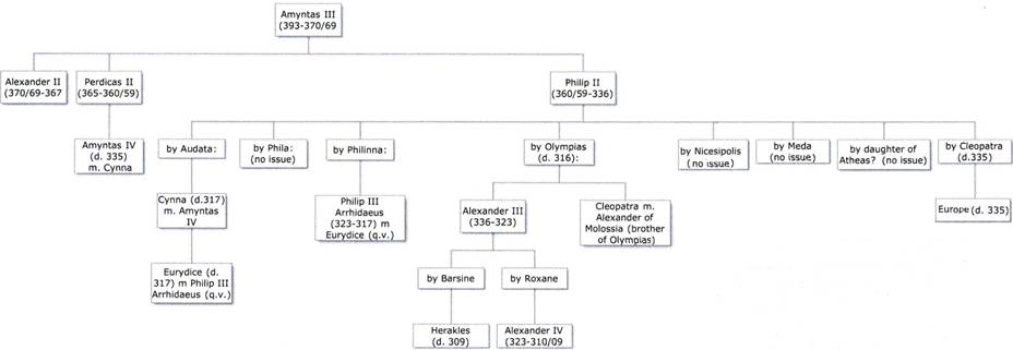
Argead family tree.
2. AIM
The aim of this paper is not to press the claims of Philip II and Cleopatra, but to draw attention to the flaws in those for Philip III Arrhidaios and Eurydice. Their case has received a lot of support before and after Bartsiokas' article (1) was published, not least by Eugene Borza and Olga Palagia. In 2007 they revived it (3).
The following year Miltiades Hatzopoulos published an 'evaluation of the unending controversy about the identity of the occupants of Tomb II' (4). It is an account of opposing claims made in the thirty years between the discovery of the tomb in November 1977 and November 2007. In it he drew attention to Bartsiokas' reliance on the pro-Arrhidaios publications of Borza and Palagia in particular, which led to conclusions reflecting 'the author's [Bartsiokas'] incomplete and partial documentation'.
We have also expressed our disagreement with Bartsiokas (1), with more emphasis on the historical context, in the festschrift in honour of the distinguished Greek historian John Davies (5).
3. OSTEOLOGY
3.1. The notch on the superior margin of the right orbit 1
In 1984 we drew attention to a notch on the superior margin of the right orbit and an associated pimple on its inner surface (6, 61-62). Using macrophotography Bartsiokas explained their existence as follows. '[T]he “pimple” anatomically corresponds to the bony protuberance of the supraorbital notch and therefore does not constitute evidence of bone remodeling or callus formation … Similarly, the “notch” is identified with what is anatomically termed the frontal notch (Figs. 1 and 2) and bears no evidence of healing or callus formation as would be expected in a notch made by an arrowhead.' (1, 512).
Our suggestion was tentative and we see no reason for departing from our cautious approach to the cause of these two features on a very small area on the upper facial skeleton which was also damaged. 'Even if our suggestion that the upper margins of the eye sockets are asymmetrical is accepted, we still have to admit that trauma need not necessarily have been entirely responsible for the apparently exaggerated notch on the right.' (6, 62). See our Fig. 2.
Figure 2
Vergina Tomb II main chamber male. Exaggerated notch in the superior margin of the right orbit (arrowed).
3.2. Facial asymmetry 1: right zygomatic bone and right maxilla
Because Bartsiokas (1) denied the existence of any arrow wound to the right side of the face he naturally dismissed what we suggested might be a healed fracture on the right zygomatic bone associated with a nick taken out of it and the right maxilla where they meet at zygomaxillare. In his words 'there is no evidence of healing at this suture'; what 'the skull shows is bone distortion owing partly to cremation and partly to a poor reconstruction of the facial skeleton'.
We reply that restoring delicate areas of the face, especially when they have been cremated, is more difficult than gluing together limb bone fragments that join perfectly. The presence of glue and consolidant here has always made study of this area difficult. See our Fig. 3. This photograph of the zygomatic bones and maxillae, taken by JHM in August 1984, shows the state of reconstruction and repair then.
Figure 3
Vergina Tomb II main chamber male. Zygomatic bones and anterior aspect of his maxillae. Note nick at right zygomaxillare (arrowed) and the asymmetry in the curvature of the lateral walls of his maxillae (arrowed below).
3.3. Facial asymmetry 2: lateral walls of the right and left maxillary sinuses
In 1984 JHM wrote 'Possibly connected with this [trace of a healed fracture] is the asymmetry in the curvature of the lateral wall of the maxilla as it curves downwards from its junction with the zygomatic bone (zygomaxillare) to the gingival margin of the upper molar teeth. On the left the outline is normal; on the right it looks decidedly abnormal.' (6, 63). In 1985 he added 'Words like shorter, straighter and more angled come to mind to describe it.' (7, 5).
Bartsiokas (1) disagreed: 'Similarly, the reported gross asymmetry between the lateral walls of the right and left maxillary sinuses … is a result of this poor reconstruction (Fig. 3). Our reply is that this reconstruction took place above the lateral wall of each maxillary sinus and would not have affected the specific region we claim shows asymmetry.
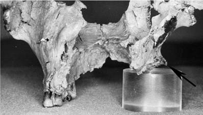
In a lecture JHM gave in Athens in 1989 he referred to this region as follows. 'On the next [two] slides are his cheek bones and maxillae — delicate parts of our anatomy at the best of times. This piece is so complete that you can easily make out a gross asymmetry in the contour of the lateral wall of the right maxillary sinus. On [his] left … it is normal. It is even more striking when viewed from behind. Note too that I had to jack the right side up on perspex disks to orientate the hard palate along its normal horizontal plane.' (8, 277). We still contend that this asymmetry is real. See our Figs 3, 4 and 5.
Figure 4
Vergina Tomb II main chamber male. Posterior aspect of his maxillae. Note the asymmetry in the curvature of their lateral walls (arrowed).
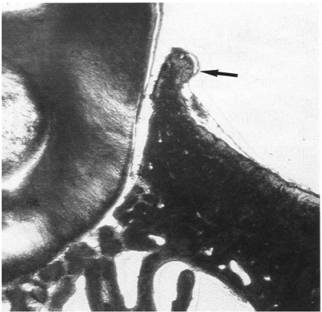
Figure 5
Vergina Tomb II main chamber male. Resorption and lipping (arrowed) of right alveolar margin due to chronic infective periodontal disease? Note again the asymmetry in the curvature of their lateral walls.
3.4. The presence of osteophytes and alveolar resorption
JHM continued 'It is clear that, in addition to losing the normal swooping curve of the maxillary wall, he had lost a lot of alveolar bone by resorption on his right side. I should stress that this condition was caused neither by post mortem damage, nor by the action of fire. The alveolar margin was in no way damaged, and the presence of osteophytes — little bony growths — along its buccal margin helps us to establish that its plane in life, and at death, was as found.' (8, 277).
Bartsiokas (1) disagreed. The presence of exostoses, as he preferred to call them, — incorrectly 2 — and alveolar resorption as a result of possible periodontal disease 'is too limited to account for the seeming asymmetry.'
We believe there is ample evidence of alveolar resorption on the right side. The need to jack up the right alveolar margin to orientate the hard palate along its horizontal plane has already been mentioned. This and an osteophyte are easily seen in a close-up image of the region (Fig. 5). The latter is very similar to one illustrated in an authoritative atlas of forensic dentistry (9) reproduced in Fig. 6. Nor is there any evidence of 'poor reconstruction' of the hard palate and gingival margin of this man. Fig. 7 is a photograph taken by JHM in 1983 of the remnants of his upper dentition and hard palate. Despite some fire-induced warping laterally, it is clear that all sixteen tooth sockets are undamaged and the parabolic curvature of the dental arcade had been preserved. See too Plate 53(a) in (10). As JHM described it in 1985, 'the alveolar margin is markedly resorbed. It is still horizontal but the amount of bone remaining has been diminished vertically so that the sockets for all three molar teeth … are considerably shallower than those on the left. That this resorption is natural and … not fire-created is indicated by … a cluster of osteophytes along the buccal rim of all three molar sockets.' (7, 5).
Figure 6
Resorption and lipping of alveolar margin due to chronic infective periodontal disease. Fig. 284 in Reference 9 (Whittaker and MacDonald 1989). Reproduced with permission from Elsevier Limited.
Figure 7
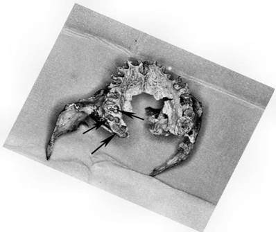
Vergina Tomb II main chamber male. Occlusal view of remnants of his maxillary dentition and hard palate. Resorbed and shallow sockets for all three right molar teeth arrowed.
4. TAPHONOMY
4.1. The difference between bones burned green/fleshed and those burned dry/degreased
Bartsiokas made it all sound so simple. 'It has been suggested that after Arrhidaeus' assassination and burial by Olympias in 317 B.C., Cassander exhumed, cremated, and reburied Arrhidaeus the following year; that is, about 6 months after his death …… So the critical question that would determine the identity of the cremains is whether there is any way of determining from the bones themselves whether they were cremated with flesh around them or cremated dry (degreased) after the flesh had been decomposed by burial.' (1, 513). He opted — incorrectly — for the latter.
Bones are often referred to as 'green' when they have been burned either as part of a whole flesh-clad body or as part of a decomposed and skeletonised one. 'Dry' bones are completely degreased, either naturally or chemically. Examples of dry bones are found in dissecting rooms, museums and archaeology departments. As Bartsiokas correctly observed, the fracture lines on bones burned 'green' differ from those burned 'dry'.
'Long bones cremated fleshed [our bold] are fragmentary with marked warping; they assume a white, blue, and gray color and present frequent and parallel-sided transverse fractures that are either curved (thumbnail) or serrated.' (1, 513).
'Long bones cremated dry [our bold] are nearly intact in size and form and show negligible warping; they assume a light brown color and present infrequent and straight transverse fractures … or step fractures.' (1, 513).
Examples of bones cremated fleshed and dry are illustrated and described in Figs 8-11. Figs 8 and 10 are reproduced, with permission, from (11), where they appeared as Figures 50 and 51.
In Table 1 we have set out T D Stewart's excellent summary of Raymond Baby's cremation experiment performed to demonstrate the differences between bones burned fleshed and dry (with our emphasis).
Table 1
Results of Raymond Baby's cremation experiment in 1950 summarised by T D Stewart in (12) and tabulated by JHM.
| Authority | Material | Burning | Result |
|---|---|---|---|
| (13) and (14) | 1. A whole, unembalmed, fleshed body from the Department of Anatomy at Western Reserve University. | Three-jet furnace; preheated to 816° C; remains pushed into three separate areas of it; gas increased and temperature raised to 1149° C; maintained for 2½ hours, completely reducing the remains. Burnt residue carefully removed the following day. | Exhibited deep checking, warping, reduction of compact bone, and transverse and diagonal fractures ('hinging'). There was absolutely no observable difference between the two types of green bone, both fleshed and defleshed. |
| 2. An almost completely defleshed cadaver from the dissecting room | Ditto | Ditto | |
| 3. Dry bone (macerated, degreased and in use by students for some ten years) | Ditto | Remained nearly intact, with little or no reduction in size or alteration in form. Exhibited superficial checking, fine longitudinal striae, deep longitudinal fracturing, or splintering, and no warping. |
Figure 8
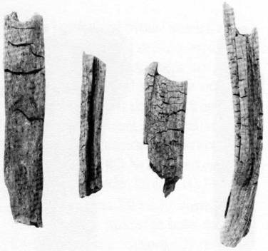
Cremated bones showing crackling and longitudinal splitting, indicating that they were dry when burned. Fig. 50 in Reference 11 (Ubelaker 1999). Reproduced with permission from Taraxacum.
Figure 9
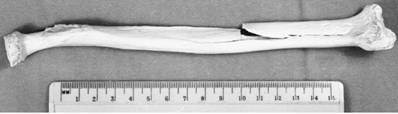
Right radius, showing longitudinal splitting and minimal warping, from JHM's dry burning experiment.
Figure 10
Cremated bones with transverse fracture lines, irregular lengthwise splitting, and marked warping, indicating that cremation was done when the bones were 'green' or covered with flesh. Fig. 51 in Reference 11 (Ubelaker 1999). Reproduced with permission from Taraxacum.
4.2. On what evidence did Bartsiokas base his assertion that the male skeleton from the main chamber in Tomb II was cremated dry?
4.2.1. Long bones
Bartsiokas continued 'As can be observed from the long bones of the male skeleton, the preservation of the bones is excellent, with minimal warping and transverse cracking that is straight (Fig. 5). The skeleton is almost complete, and light brown is the dominant color of the bones. Only the left proximal ulna presents some curved transverse fractures, probably the result of insufficient decomposition in this area. The right ulna is nearly perfect, with a longitudinal crack. This type of preservation of the male skeleton shows that most of the bones were dry when cremated; that is, they were buried for some time before they were cremated. This is consistent with the taphonomic history of inhumation, cremation, and reinterment that only the bones of Arrhidaeus underwent, as already mentioned.' (1, 513-514).
To refer to 'insufficient decomposition' on 'the left proximal ulna' displays a lack of understanding of the process and its timescale. Did Bartsiokas expect his readers to accept that it was possible for a few centimetres of this bone to have retained putrefying soft tissue while the rest of it desiccated completely in a matter of months?
In the legend to his Fig. 5 Bartsiokas described the left tibia as 'A typical example of a long bone from the male skeleton: nearly intact, with minimal warping and a step transverse fracture, which are all evidence of a dry bone cremation consistent with the taphonomic history of Arrhidaeus.' (1, 514).
Of more interest are fractures at the proximal end of the bone. On JHM's image of the paired bones (Fig.12) transverse fracture lines are visible on the medial surface of the upper shaft and the tibial tuberosity has been damaged, seemingly avulsed forwards. The tibial tuberosity is the site of insertion of the patellar ligament and ultimately of the quadriceps femoris muscle. Were the fracture lines and avulsion caused when quadriceps femoris, one of the most powerful muscles in the human body, shrank during burning? Damage of this kind is discussed and illustrated by Symes et al. in (15) (See especially Figure 2.1 and Plate 1).
The two-dimensional images of these tibiae, here and in (1), do not and cannot show the degree of warping. When the skeleton was photographed from different angles, as in Figs 13-15, this man's femora, tibiae, right humerus and left ulna in particular were clearly seen to be very warped. Plate 54b in (10) also depicts the femora as warped.
We believe that Bartsiokas' claim of 'minimal warping' does not apply to the long bones of this skeleton. If warping is a major feature of bones burned fleshed (see Table 1), the distorted shape of many of them indicates that this skeleton was cremated fleshed.
Figure 11
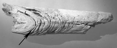
Lefkandi Toumba Tomb 79. Femoral fragment showing curved transverse fractures: classic signs of its having been burned 'green' with flesh on. Photograph courtesy of Professor Irini Lemos.
Figure 12
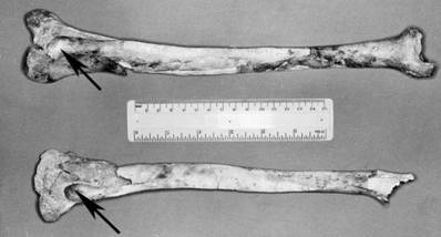
Almost complete tibiae of Vergina Tomb II main chamber male. Note symmetrical fractures to proximal ends (arrowed).
Figure 13
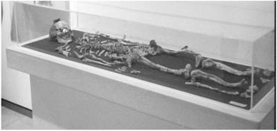
Skeleton of Vergina Tomb II main chamber male, on display in the Archaeological Museum, Thessaloniki, September 1990. Note warping of femora and tibiae, on right of image.
Figure 14
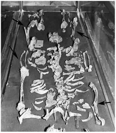
Lower part of the skeleton of Vergina Tomb II main chamber male in store in the Archaeological Museum, Thessaloniki, July 1983. Warped right humerus, left ulna and both femora arrowed.
Figure 15
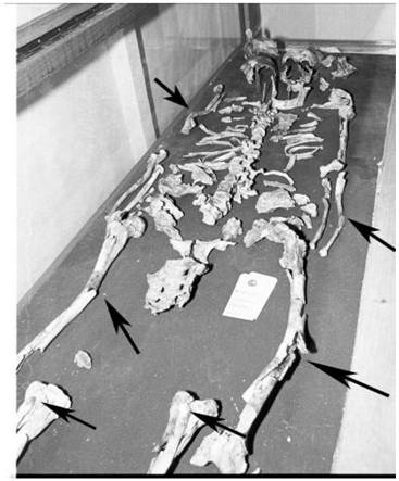
Part of the skeleton of Vergina Tomb II main chamber male in store in the Archaeological Museum, Thessaloniki, July 1983. Warped right humerus, left ulna and both femora and tibiae arrowed.
4.2.2. Cranium
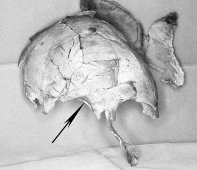
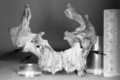
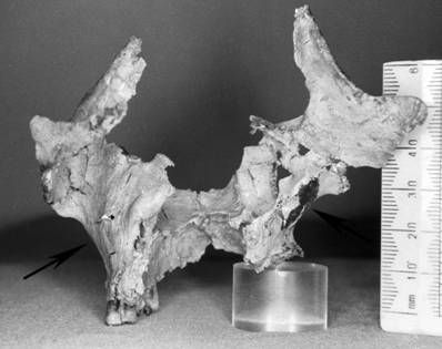
In Bartsiokas' opinion 'it is not as yet clear what happens to the flat bones.' (1, 513). As far as the cranium is concerned, we know. They warp. The most striking evidence is a flange of his left parietal bone that had been bent outwards along the natural line of the coronal suture by the heat of the fire (Fig. 16). This flange is loose. Rough handling would have detached it long ago. Another cranial vault fragment comprising much of the occipital bone and part of the left parietal offers further proof (Fig. 17). In this figure the internal surface of the occipital bone is in focus. The blurred image at the top is another flange of the left parietal bone that had become flattened and now points forward. In Fig. 18 the external surface of this fragment is viewed from the right and shows another portion of the left parietal pointing outwards and backwards.
Figure 16
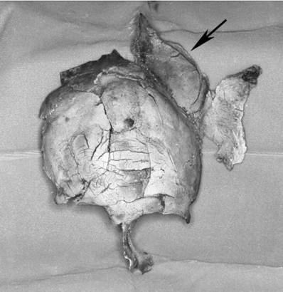
Vergina Tomb II main chamber male. Flange of left parietal bone hinged outwards along coronal suture during burning (arrowed).
Figure 17
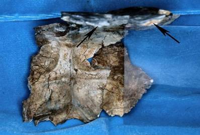
Vergina Tomb II main chamber male. Internal aspect of occipital bone with flange of the left parietal bone — out of focus at top right (arrowed) — warped out of normal alignment during burning and pointing forward. The outline of a Perspex block used to support it is visible on the right.
Figure 18
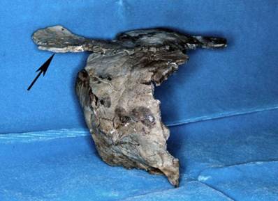
Vergina Tomb II main chamber male. External aspect of occipital bone viewed from the right and showing another portion of the left parietal bone (arrowed) pointing outwards and backwards.
Did this massive distortion of the left side of the cranial vault occur when the brain, blood and cerebrospinal fluid boiled and the skull exploded? Some authorities deny that skulls ever explode (16). Others, cited by Fairgrieve in (17), believe they do. There is abundant anecdotal evidence on the Internet. Even if the skull did not explode, warping and distortion on this scale points to fleshed cremation rather than dry, as does the wide variation in colour observed on the cranial fragments. They are also very different from the residue of an experiment conducted by JHM, in which a dry cranium was burned for seven hours at 900o C in an electric kiln, a temperature that took five hours to reach. As a result of exposure to this high temperature for so long the anterior ends of each parietal separated from the frontal bone and bent inwards (Fig. 19).
Figure 19
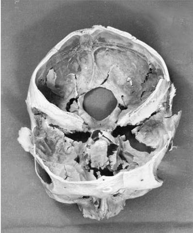
Implosion of parietal bones on a cranium from JHM's dry burning experiment.
4.2.3. Conclusion to 4
We believe that the man in the main chamber of Tomb II was burned as a fleshed cadaver on the evidence of the widespread warping and distortion of the bones of his cranial vault and at least six long limb bones: his right humerus, left ulna, both femora and both tibiae.
5. TAPHONOMIC IRONY: WHY ARRHIDAIOS COULD NOT HAVE BEEN BURIED IN TOMB II
There is another reason why the man in the main chamber of Tomb II, if he was Philip III Arrhidaios, could not have been burned dry. He was murdered in the autumn of 317 BC and buried early in 316 or possibly in the winter/spring of 316-15. See Section 9.1.2. We do not know the fate of his body during the intervening months.
One of us (HW) is a forensic pathologist, with wide experience of examining exhumed human cadavers and partially skeletonised remains discovered above ground. In his opinion, Arrhidaios' body would still have had putrefying skin and muscle attached to his limb bones, and rotting viscera filling his thoracic, abdominal and pelvic cavities after even seventeen months in the ground. It would not have become a dry and degreased skeleton.
As evidence of the resilience of such material, we cite the remains of a murder victim from the south west of England whose body had been stored for twenty months before being deposited on moorland for up to a further ten. When they were discovered they were badly decomposed. Even after maceration by enclosed boiling for several hours, his skeleton was not dessicated and traces of soft tissue still adhered to it. An example is illustrated in Fig. 20.
Figure 20
Traces of soft tissue (arrowed) still adhering to the first cervical — atlas — vertebra of a 31±5 year-old man after maceration. He had remained unburied for 30 months. Note developmental abnormality: spina bifida atlantis. Photograph by University of Bristol Department of Anatomy Digital Media Imaging Service.
If Bartsiokas is right and the bones in the gold casket were burned dry, they could never have belonged to Philip III Arrhidaios because it would have taken them several years to achieve the state of dryness to produce the fracture lines classic of dry cremation. The literature on decomposition is extensive. References (18) and (19) are useful sources.
This is hypothetical. We have already demonstrated that the bones from the main chamber were burned fleshed. It is also likely that the fleshed body of Arrhidaios was cremated soon after death on ethical grounds. For ordinary Macedonians burying him and exhuming him for cremation and reburial later would have been repugnant. 'In many ancient Greek sacred laws, every human corpse is considered a significant source of pollution' (20). See Section 9.1.2. We happen to have a description of the reaction of battle-hardened soldiers to rotting corpses at the time. To those beleaguered in Pydna during Cassander's siege in 316, the sight and stench — as recorded by Diodorus Siculus (Library of World History 19.49.4) — was abominable and unbearable.
6. THE REST OF THE FAMILY: EURYDICE AND KYNNA
Bartsiokas' study concerned the skeletal remains from the main chamber of Tomb II. He makes one reference to Philip III Arrhidaios' wife Eurydice, in the context of our previous work. Eurydice plays an important part in this sad story. Indeed it is incomplete without her. We have already mentioned that she was forced to commit suicide in 317 BC, and was buried with Arrhidaios and her mother Kynna in a new tomb.
Although he does not say so in his paper, we assume that Bartsiokas would accept the cremains in the smaller gold casket in the antechamber to Tomb II as Eurydice's. They have been described by Xirotiris and Langenscheidt in (10) and JHM in (8). These authors agree that they belonged to a woman aged between 20 and 30: 'about 25 years, more likely not younger than 20 or older than 30 years.' (10, 155-156).
Bartsiokas should have been aware of the very great quantitative difference between the two sets of cremains. The man's comprised a virtually complete skeleton with pieces ranging from whole limb bones to small ones such as undamaged cervical vertebrae. The woman's comprised 1312 g of modest bone fragments, not dissimilar to many other ancient Greek cremations examined by JHM (21), (22), (23). A representative sample is reproduced in Fig. 21.
We have not been able to find a published account of the weight of the man's cremains. Some idea may be obtained from a collection of miscellaneous fragments not on display, which JHM examined and weighed in May 1990. If, at 379.13 g, they represented 10% of the weight of the whole skeleton (3791.3 g), the latter score is close to the top of the range recorded by other investigators: 1600-3600 g (24); 2200-3750 g (25); ♂ 3379.77 g, ♀ 2350.17 g (26). If they represented 15%, the skeleton would have weighed 2527.53 g, well inside these ranges.
In a study of modern cremations in Florida (27), Warren and Maples found that 'The average cremains weight for the fully developed adults (n = 91) was 2430 g and ranged from 876 g to 3784 g. Male and female means were separated by 1053 g, but there was considerable overlap in the distribution. All cremains weights above 2750 g were male and all cremains weights below 1887 g were female.'
This disparity in the weights of these two cremated skeletons is perplexing. However it is not difficult to explain. In 1984 JHM wrote 'the skeleton from Tomb II shows every sign of having been burnt in some sort of enclosed chamber' (6, 77). This tentative suggestion was subsequently borne out by Dr Angeliki Kottaridi's study of the pyre site (28).
Figure 21
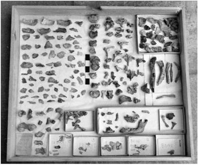
Vergina Tomb II antechamber female. One of the two trays containing her cremated remains. Length of scale bar 2.5 cm.
Dr Kottaridi's analysis of the finds from the pyre site revealed:
- Several hundred unfired mud bricks, used for the socle on which an ornate chryselephantine couch and the body were placed; and also for the construction of a small building in which they were burned, complete with a lion head door knocker.
- Weapons and arms, funerary wreaths, a wooden box for clothes, fibulae and carbonised fragments of finely woven cloth.
- A bronze libation jug and smashed plates for food offerings.
- Grape pips and almonds, and containers for aromatic oils.
- Burned bones of lambs, kids and horses.
- Harness probably intended for young race horses.
As she observed, even if the tomb had been looted, the impressive wealth of the offerings would have been interpreted as intended for an exceptional recipient. Philip III Arrhidaios would not immediately have come to mind.
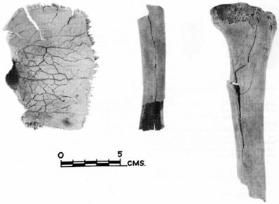
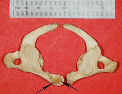
We assume that the woman from the antechamber was cremated on an open pyre. That she was burned fleshed is not in doubt. The neck and proximal shaft of each femur, especially the left, show curved transverse fractures (Fig. 22); as do several smaller limb bone shaft fragments (Fig. 23). For Bartsiokas this would presumably disqualify her from being identified as Arrhidaios' wife Eurydice as he believed her husband was cremated dry (1).
Figure 22
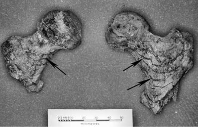
Vergina Tomb II antechamber female. Femoral neck and proximal shaft fragments showing curved transverse fractures (arrowed) that indicate they were burned fleshed.
Figure 23
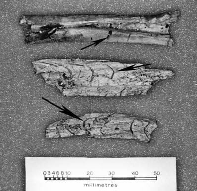
Vergina Tomb II antechamber female. Limb bone shaft fragments showing curved and straight transverse fractures (arrowed) that indicate they were burned fleshed.
There is another reason why the woman in the antechamber might not have been Eurydice: her age. According to RLF, in Section 9.1.3, she was between 18 and 19 'and some months' in October 317. There are no features on the bones from the antechamber that would permit an estimate as low as this.
7. THE CASE FOR ARRHIDAIOS AND EURYDICE GROWS WEAKER
7.1. The absence of Kynna
The argument in favour of Arrhidaios and Eurydice as the occupants of Tomb II is further undermined by the absence from Tomb II of the remains of a third person. Both Diodorus Siculus (19.52.5) and Diyllos (29) imply that Kynna, Eurydice's mother, was buried alongside her daughter and son-in-law Arrhidaios. Presumably Kynna's ashes were already in an urn as she was executed in Asia by Alcetas in c. 322 BC (8, n.30). See too Section 9.1.2.
7.2. The burials of Arrhidaios, Eurydice and Kynna: reasonable expectations
From the historical account of their deaths and committals, one might expect:
- The cremated remains of Arrhidaios and Eurydice to have been qualitatively and quantitatively similar as regards fragment size and the amount of bone placed in each one's container.
- Their containers to have been interred in the same chamber.
- A third to be present near the other two holding the bones of Kynna.
- Or one receptacle containing the remains of Arrhidaios and Eurydice and another Kynna's. The famous Derveni Krater, from the high status Tomb Beta at Derveni, contained the cremated remains of a man and a woman (21).
8. SUMMARY AND CONCLUSION
We answer Bartsiokas' criticism of our early work on a very small area on the upper facial skeleton of the man in the main chamber of Tomb II at Vergina. We also maintain that there is ample evidence of asymmetry in the lateral wall of the right maxillary sinus; and of alveolar resorption on the right side of the maxilla that may have resulted from periodontal disease. We have demonstrated that he was mistaken in claiming the skeleton was burnt dry rather than fleshed. He understood the differences in appearance between bones burned fleshed and those burned dry. However he seems not to have understood how long it takes a fleshed cadaver to become a completely dry, degreased skeleton. We have drawn attention to the consequences for the pro-Arrhidaios supporters of this misunderstanding. Finally we do not believe that the qualitative and quantitative differences between the two collections of bones in Tomb II, their committal in separate caskets and chambers, and the absence of a third individual fit the descriptions of the funeral Cassander laid on at Aigai for an undistinguished Argead king, his wife and his mother-in-law.
9. PERSPECTIVES
9.1. Vergina: a short historical perspective
9.1.1. Introduction
This is not the place to answer the pro-Philip III Arrhidaios points made by Borza and Palagia in 2007 (3). RLF will develop this in a paper currently in progress. Instead the aim of this short historical perspective is to revisit the evidence for the dating of various events which are believed to be relevant by the Philip III Arrhidaios supporters but which rule him and Eurydice out as candidates for Tomb II.
9.1.2. The chronology of the burial and reburial of Philip III Arrhidaios and Eurydice
In October 317, as guaranteed by Diodorus (19.11.5), Philip III Arrhidaios and Eurydice were murdered in Macedon on Olympias's orders. Between four and 17 months later — see below — they were reburied at Aigai by Cassander, together with Kynna.
Kynna had been murdered in Asia some four or five years earlier and presumably had been cremated and preserved as bones, like other dead dignitaries of the age of Alexander the Great's early Successors. A classic example is general Craterus' cremated bones being given to his wife for burial several years later (Diodorus 19.59.3). The dead Eumenes received similar treatment (Diodorus 19.44.2).
The length of time between Philip and Eurydice's deaths and their subsequent reburial by Cassander is usually reckoned to be up to four months. However, Anson (30) has now made a sustainable case for their honorary funeral being as late as winter-spring 315 BC, up to 17 months later. His longer chronology seems not impossible, although it has not been tested fully by counter-arguments as yet. It is independently suggested by Boiy (31, 113-116).
It is unlikely that the bodies of Philip and Eurydice were ever exhumed as rotting corpses for subsequent cremation at Aigai or elsewhere. It is unparalleled and incredible that corpses recovered in this way from their initial burial would have been secondarily cremated at their new resting-place. Their rehandling would have been seriously polluting for participants and grotesquely contrary to Greek beliefs about contact with deposed corpses. They were transferred, surely, as already-cremated skeletons, the result of a rite paid to them by well-wishers after their deaths. They could not therefore have been re-cremated on the pyre inside the special funerary house of wood, attested beside Vergina Tomb II. Their cremation had already occurred months before, like the cremations of other distinguished dead, whether Eumenes or Craterus, or the princely youth (possibly Alexander IV) in Tomb 1V.
9.1.3. The chronology of the birth of Eurydice
We know that Eurydice, Philip III's bride, died in October 317, but when was she born? Her mother Kynna was daughter of Philip II and the Illyrian Audata, who was married to him in 359-8 BC following his first victory over an Illyrian army. Kynna cannot have been born before 358 and may have been a year or more younger. When did she, in turn, marry? This is a complicated story. Fig. 1 will make it easier to follow.
Kynna's first — and only — husband was Philip II's nephew Amyntas. Some think he was briefly king as a boy before Philip II, and thus he is known as Amyntas IV to modern scholars. According to Polyaenus (Stratagems 8.60), writing in the early 160s AD but using much earlier sources, her marriage to Amyntas ended 'swiftly'. Polyaenus uses his sources to emphasise the independent spirit of Kynna. Her 'swift' ending of this marriage, and her refusal to contract another, are integral to his section about her and are based on evidence about her life.
Amyntas had certainly died by summer 335 BC, on the evidence of Arrian (Anabasis 1.5.4), who refers to Alexander the Great's offer to marry her to Langarus the Agrianian, made to him in summer 335 BC and to be acted on when Langarus had come to Pella. In fact he never came and there was no marriage because Langarus died first of sickness. Plutarch (Moralia 327C) claims rhetorically, but plausibly, that Amyntas was a focus of royal opposition to Alexander's accession in autumn 336 BC after Philip II's assassination. It is therefore likely that Amyntas was put to death in the usual purging of rival kin which followed a Macedonian king's accession
With Amyntas dead by spring-summer 335, his marriage with Kynna cannot have begun earlier than, say, 337 to be as 'swiftly' ended as the story requires, and 336 is perfectly possible too. At that time the bride, Kynna, was up to 21 years old, fitting with such comparable evidence as we have for other Macedonian royal brides, as collected by Greenwalt (32, 94-95).
The wedding of a daughter to a royal nephew was an important match within Philip II's family and surely one which he would have personally brought about and encouraged. It would be an occasion which he would most probably wish to attend in person, as he was to attend the similar marriage of his daughter Cleopatra in autumn 336 at Aigai. Philip's own travels are then a limiting factor too. They took him away from Macedon from winter 342-1 until late summer 339 and then in the autumn down into central Greece, where he stayed to fight at Chaeronea in late August 338, and then on to Sparta that autumn in the aftermath. The wedding, if in Philip's presence at court, cannot have occurred in early 337 because Philip was still in the south, organising his Hellenic League meetings at Corinth. From this angle too 337-6 BC is the likeliest date for a doubly royal wedding in Macedon to be celebrated in his presence. The chronology of Philip's movements in these years is not controversial and the available facts are correctly set out by Hammond and Griffith (33, 725-726).
Seeing the dangers here for supporters of the Philip III theory, Adams (34)[especially 72, n.60], tried to claim that Kynna could have married Amyntas as early as 342 BC so that Eurydice could be about 24 when she died. This evasion is not possible. Amyntas was still alive in 336 and a six-year marriage with him would not permit the gloss in Polyaenus that Kynna ended it 'swiftly'. She had not, of course, divorced Amyntas on her own initiative before 336 BC: the marriage ended 'swiftly', but only because of his death.
As a young girl of nineteen and some months, or perhaps eighteen going on nineteen (if born, say, in late 336 or very early 335) Eurydice died too young in October 317 to be the lady in Tomb II.
9.2. People and archaeology
9.2.1. Background to this paper
The specific arguments made above should be placed in a wider context. They arose from Richard Neave's reconstruction of the head of the male occupant of Tomb 2 at Vergina (6).
9.2.2. History of facial reconstruction
Facial reconstruction is not a new technique. It began with the plaster faces applied to skulls in Neolithic Syria and Jericho (35), (36), (37). However, scientific attempts to reconstruct the face of a particular individual (as opposed to death masks and anatomical models) really started with the work of Wilhelm His (1831-1904) towards the end of the nineteenth century, and the technique was brought into the twentieth century by the Russian palaeontologist Mikhail Gerasimov (1907-1970). The later twentieth century brought developments that made it both an essential tool in forensic science and an important element in archaeological research and museum displays. The team led by Richard Neave at the Unit of Art in Medicine in the University of Manchester played a very significant role in these developments (37) (38).
9.2.3. Impact of facial reconstruction
Encouraged by the current interest in local history and in genealogy, every museum and every archaeological television programme now has its facial reconstruction. Whether carried out by building up the facial muscles in clay or wax on a cast of the skull or by using various forms of computer technology, the underlying tenet is that the bony skull forms the armature for the fleshy face and thus dictates its form. Data collected since the late nineteenth century now provide full information for the varying flesh thicknesses of different age, sex and ethnic origin.
However, the technique has much greater potential than mere display. To achieve it requires: (1) detailed knowledge of anatomy (cranial and post-cranial); (2) familiarity with the circumstances of burial and the history of the individual(s); and (3) a thorough understanding of the three-dimensional relationship of the often distorted pieces of a fragmentary or incomplete skeleton. All of this can only be achieved by studying the actual remains and not photographs, however good they may be. Done properly therefore, it becomes a three-dimensional report that can convey information in a way that mere print (even e-print) can never do.
If nothing else, the case of the skull in Tomb II at Vergina shows the scope and potential for cross-disciplinary work in facial reconstruction. Work at Manchester has involved anatomists, archaeologists, biologists, epigraphists, geneticists, historians, make-up artists, pathologists, policemen, radiographers, surgeons and so on. Its accuracy is guaranteed by controls, both basic 'before and after' photographs of cadavers in the anatomy school and reconstructions of living people made on replica skulls derived from CAT-scans.
Because it is a purely objective method, based on the information provided by the skull itself, facial reconstruction can be used to trace kinship based on the facial similarity that often exists between members of the same family. We have used it to trace dynastic links between burials in Bronze Age Mycenae. As they were prehistoric, no written sources exist to guide us. However the results can often be controlled and confirmed by analysing their DNA (37), (39), (40), (41).
9.2.4. People matter in archaeology
Sir Mortimer Wheeler insisted that 'the archaeologist is digging up, not things, but people.' (42)(page 13). He wished us not to become obsessed with potsherds and scraps of metal for their own sake, but to remember the people who made and used them. This is what we are doing now. The poet Seamus Heaney put it very well in 'The Grauballe Man', a poem about bog bodies: 'Who will say 'corpse' to his vivid cast?' (43).
However, this interest in humanity has raised another important question. There are archaeologists and museum curators who are becoming increasingly concerned at the ethics of working with human remains and of putting them on display. For a balanced view see (44), (45)(pages 17-37) and (46).
Unlike the subjects displayed in Gunther von Hagens' 'Body Worlds' exhibitions and in his Institute for Plastination in Heidelberg (www.bodyworlds.com), we cannot consult the people whose remains we reconstruct. Nevertheless we believe that what we do causes no offence either to the dead whose stories are revealed or to students of our results in the search for scientific and historical truth. This is not to say that there are not those who have made their views very clear. A notable example is William Shakespeare, whose tomb carries the well-known minatory inscription:
Good frend for Iesus sake forbeare
to dig the dust encloased heare:
Blese be ye man yt spares thes stones,
And curst be he yt moves my bones.
10. ACKNOWLEDGEMENTS
We thank Elsevier Limited for permission to reproduce Fig. 284 in (9) as our Figure 6; Taraxacum for permission to reproduce Figures 50 and 51 in (11) as our Figures 8 and 10; Professor Irini Lemos for Figure 11; and Professor H A Waldron and Professor I A Silver for advice on anatomical terminology. We are especially grateful to Dr Angeliki Kottaridi and the late Dr Julia Vokotopoulou and Professor Manolis Andronicos for their encouragement and permission to study these important human remains.
CONFLICT OF INTEREST
The authors have declared that no conflict of interest exists.
References
1. Bartsiokas A. The eye injury of King Philip II and the skeletal evidence from the Royal Tomb II at Vergina. Science. 2000;288:511-14
2. Andronicos M. Vergina: the royal tombs and the ancient city. Athens: Ekdotike Athenon. 1984
3. Borza EN, Palagia O. The chronology of the Macedonian royal tombs at Vergina. Jahrbuch des Deutschen Archäologischen Instituts. 2007;122:81-125
4. Hatzopoulos M. The burial of the dead (at Vergina) or the unending controversy on the identity of the occupants of Tomb II. Tekmeria. 2008;9:91-118
5. Prag AJNW, Musgrave JH, Neave RAH. A twenty-first century Philippic. In: (ed.) Oliver GJ, Archibald Z. The Power of the individual in ancient Greece. Essays in honour of Professor JK Davies Stuttgart: Franz Steiner publication expected in 2010-11
6. Prag AJNW, Musgrave JH, Neave RAH. The skull from Tomb II at Vergina: King Philip II of Macedon. Journal of Hellenic Studies. 1984;104:60-78
7. Musgrave JH. The skull of Philip II of Macedon. In: (ed.) Lisney SJW, Matthews B. Current topics in oral biology. Bristol: University of Bristol Press. 1985:1-16
8. Musgrave JH. Dust and damn'd oblivion: a study of cremation in ancient Greece. Annual of the British School at Athens. 1990;85:271-99
9. Whittaker DK, MacDonald DG. A colour atlas of forensic dentistry. London: Wolfe Medical Publications. 1989
10. Xirotiris NI, Langenscheidt F. The cremations from the royal Macedonian tombs of Vergina. Archaiologiki Ephimeris. 1981:142-60
11. Ubelaker DH. Human skeletal remains: excavation, analysis, interpretation, 3rd ed. Washington: Taraxacum. 1999
12. Stewart TD. Essentials of forensic anthropology. Springfield: Charles C Thomas. 1979
13. Baby RS. Hopewell cremation practices. Ohio Historical Society, Papers in Archaeology, No. 1. Columbus, Ohio. 1954
14. Baby RS. Personal communication. Stewart TD. Essentials of forensic anthropology. Springfield: Charles C Thomas. 1979:60-61
15. Symes SA, Rainwater CW, Chapman EN, Gipson DR, Piper AL. Patterned thermal destruction of human remains in a forensic setting. In: (ed.) Schmidt CW, Symes SA. The analysis of burned human remains. London: Academic Press. 2008:15-54
16. Pope EJ, Smith OC. Identification of traumatic injury in burned cranial bone: an experimental approach. Journal of Forensic Sciences. 2004;49:431-40
17. Fairgrieve SI. Forensic cremation recovery and analysis. Boca Raton: CRC Press. 2008
18. Mant AK. Knowledge acquired from post-War exhumations. In: (ed.) Boddington A, Garland AN, Jannaway RC. Death, decay and reconstruction. Approaches to archaeology and forensic science. Manchester: Manchester University Press. 1987:65-78
19. Fiedler S, Graw M. Decomposition of buried corpses, with special reference to the formation of adipocere. Naturwissenschaften. 2003;90:291-300
20. von Staden H. The discovery of the body: human dissection and its cultural contexts in ancient Greece. The Yale Journal of Biology and Medicine. 1992;65:223-41
21. Musgrave JH. The cremated remains from Tombs II and III at Nea Mihaniona and Tomb Beta at Derveni. Annual of the British School at Athens. 1990;85:301-25
22. Musgrave JH. The human bones. In: (ed.) Coldstream JN, Catling HW. Knossos The North Cemetery: Early Greek Tombs I-IV. London: The British School at Athens (Supplementary Volume No. 28). 1996:677-702
23. Musgrave JH. Appendix A: An anthropological assessment of the cremations and inhumations from the Early Iron Age cemetery at Torone. In: (ed.) Papadopoulos J. The Early Iron Age cemetery at Torone. Monumenta Archaeologica 24. Los Angeles: UCLA Cotsen Institute of Archaeology. 2005:243-315
24. McKinley JI. Cremations: expectations, methodologies and realities. In: (ed.) Roberts CA, Lee F, Bintliff J. Burial archaeology: current research, methods and developments. Oxford: British Archaeological Reports, British Series 211. 1989:65-76
25. Holck P. Cremated bones. Oslo: Anatomical Institute, University of Oslo. 1986
26. Bass WM, Jantz RL. Cremation weights in East Tennessee. Journal of Forensic Sciences. 2004;49:901-4
27. Warren M, Maples W. The anthropometry of contemporary commercial cremation. Journal of Forensic Sciences. 1997;42:417-23
28. Kottaridi A. Vasilikes pires sti Nekropoli ton Aigon. Ancient Macedonia VI, International Symposium I. Thessaloniki: Institute for Balkan Studies. 1999:631-42
29. Diyllos. 73 F 1. In: (ed.) Jacoby F. Fragmente der griechischen Historiker. Berlin: Weidmann. 1926:131
30. Anson EM. Dating the deaths of Eumenes and Olympias. Ancient History Bulletin. 2006;20:1-8
31. Boiy T. Between high and low. A chronology of the early Hellenistic period. Frankfurt: Verlag Antike. 2007
32. Greenwalt WS. The marriageability age at the Argead court: 360-317 B.C. Classical World. 1988;82:93-7
33. Hammond NGL, Griffith GT. A History of Macedonia, Volume II. Oxford: Clarendon Press. 1976
34. Adams WL. The royal Macedonian tomb at Vergina: an historical interpretation. Ancient World. 1980;3:67-72
35. Merrony M. Plastered Syrian skulls from the dawn of civilisation. Minerva. 2007 18.1: 4-5
36. Stordeur D, Khawam R. Les crânes surmodelés de Tell Aswad (PPNB, Syrie). Premier regard sur l'ensemble, premières réflexions. Syria. 2007;84:5-32
37. Prag J, Neave R. Making faces using forensic and archaeological evidence. London: British Museum Press. 1997(reprinted with corrections 1999)
38. Wilkinson C. Forensic facial reconstruction. Cambridge: Cambridge University Press. 2004
39. Bouwman AS, Brown KA, Prag AJNW, Brown TA. Kinship between burials from Grave Circle B at Mycenae revealed by ancient DNA typing. Journal of Archaeological Science. 2008;35:2580-84
40. Bouwman AS, Brown KA, Brown TA, Chilvers EA, Arnott R, Prag AJNW. Kinship in Aegean prehistory? Ancient DNA in human bones from mainland Greece and Crete. Annual of the British School at Athens. 2009;104:291-307
41. Brown TA, Brown KA, Flaherty CE, Little LM, Prag AJNW. DNA analysis of bones from Grave Circle B at Mycenae: a first report. Annual of the British School at Athens. 2000;95:115-19
42. Wheeler M. Archaeology from the Earth. Harmondsworth: Penguin Books. 1956
43. Heaney S. New Collected Poems 1966-1987. London: Faber and Faber. 1990
44. Bienkowski P, Chapman M. Authority and decision-making over British human remains: issues and challenges. In: (ed.) Lewis ME, Clegg M. Proceedings of the ninth annual conference of the British Association for Biological Anthropology and Osteoarchaeology. British Archaeological Reports, International Series 1918. Oxford: Archaeopress. 2009:99-105
45. Roberts CA. Human remains in archaeology: a handbook. York: Council for British Archaeology. 2009
46. Williams H. Death becomes us. Minerva. 2010;21:42-5
Footnotes
1
Because it has been necessary to quote extensively from original sources in sections 3 and 4, we have decided — reluctantly — to emphasise some words to guide readers through technical aspects of the argument.
2
In the opinion of our colleague Professor H A Waldron, osteophyte 'is a much more suitable term as it implies some abnormal growth of bone whereas an exostosis is a benign overgrowth of normal bone.'
Author contact
![]() Corresponding author: Dr J H Musgrave, Centre for Comparative and Clinical Anatomy, University of Bristol, Southwell Street, Bristol, BS2 8EJ, UK Tel (UK): 0117-928-7417, E-mail: j.h.musgraveac.uk
Corresponding author: Dr J H Musgrave, Centre for Comparative and Clinical Anatomy, University of Bristol, Southwell Street, Bristol, BS2 8EJ, UK Tel (UK): 0117-928-7417, E-mail: j.h.musgraveac.uk
Citation styles
APA
Musgrave, J., Prag, A.J.N.W., Neave, R., Fox, R.L., White, H. (2010). The Occupants of Tomb II at Vergina. Why Arrhidaios and Eurydice must be excluded. International Journal of Medical Sciences, 7(6), s1-s15. https://doi.org/10.7150/ijms.7.s1.
ACS
Musgrave, J.; Prag, A.J.N.W.; Neave, R.; Fox, R.L.; White, H. The Occupants of Tomb II at Vergina. Why Arrhidaios and Eurydice must be excluded. Int. J. Med. Sci. 2010, 7 (6), s1-s15. DOI: 10.7150/ijms.7.s1.
NLM
Musgrave J, Prag AJNW, Neave R, Fox RL, White H. The Occupants of Tomb II at Vergina. Why Arrhidaios and Eurydice must be excluded. Int J Med Sci 2010; 7(6):s1-s15. doi:10.7150/ijms.7.s1. https://www.medsci.org/v07p00s1.htm
CSE
Musgrave J, Prag AJNW, Neave R, Fox RL, White H. 2010. The Occupants of Tomb II at Vergina. Why Arrhidaios and Eurydice must be excluded. Int J Med Sci. 7(6):s1-s15.
This is an open access article distributed under the terms of the Creative Commons Attribution (CC BY-NC) License. See http://ivyspring.com/terms for full terms and conditions.