Impact Factor ISSN: 1449-1907




Int J Med Sci 2026; 23(12):3678-3692. doi:10.7150/ijms.132471 This issue Cite
Review
Comparative Analysis of the Role of Reactive Oxygen Species in Endometriosis and Endometrial Cancer
Jeongmin Lee1†, Seung Geun Yeo1,2,3†, Jae Min Lee2, Hye Ok Kim1,3, Manish Kumar Singh4, Sung Soo Kim4, Tong In Oh5, Jisu Yun6, Dong Choon Park6 
1. Department of Medicine, College of Medicine, Kyung Hee University Medical Center, Seoul, Korea.
2. Department of Otorhinolaryngology Head & Neck Surgery, College of Medicine, Kyung Hee University Medical Center, Seoul, Korea.
3. Clinical Research Institute, Kyung Hee University Medical Center, Seoul 02447, Korea.
4. Department of Biochemistry and Molecular Biology, College of Medicine, Kyung Hee University, Seoul, Korea.
5. Department of Biomedical Engineering, College of Medicine, Kyung Hee University, Seoul, Korea.
6. Department of Obstetrics and Gynecology, St. Vincent's Hospital, College of Medicine, The Catholic University of Korea, Seoul, Korea.
† These authors contributed equally to this work.
Received 2026-2-1; Accepted 2026-9-4; Published 2026-11-6
Abstract
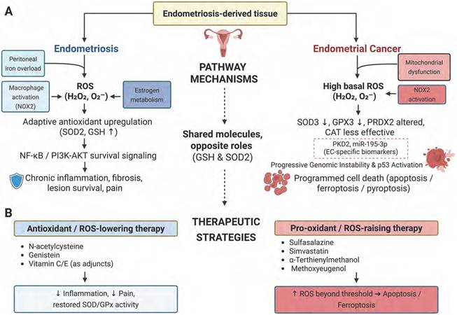
Objective: We conducted a comprehensive literature review to evaluate the roles of reactive oxygen species (ROS) and free radicals in the pathogenesis of endometriosis and endometrial cancer, which both originate from the endometrium but are clinically distinct.
Methods: A literature search employing the Cochrane Library, EMBASE, Google Scholar, PubMed, and SCOPUS databases was conducted to assess differences in the induction and role of ROS in the development of endometriosis and endometrial cancer. Given the broad mechanistic scope and the comparative nature of our analysis, this work was conducted as a narrative review to synthesize current evidence on ROS-mediated pathways in both conditions.
Results: Of the 230 articles initially screened, 76 were ultimately reviewed; 8 reported that ROS contribute to the pathogenesis of endometriosis, whereas 18 reported the involvement of ROS in the pathogenesis of endometrial cancer. Certain molecules, including hydrogen peroxide (H₂O₂), superoxide anion (O₂⁻·), lipid peroxides (LPOs), malondialdehyde (MDA), catalase (CAT), and glutathione (GSH), were commonly expressed and implicated in the pathogenesis of both endometriosis and endometrial cancer. Other factors, such as superoxide dismutase 3 (SOD3), polycystic kidney disease 2 (PKD2), heme oxygenase-1 (HO-1), p53, phosphatase and tensin homolog (PTEN), gasdermin-D (GSDMD), and the NOD-, LRR- and pyrin domain-containing protein 3 (NLRP3) inflammasome were studied exclusively in the context of endometrial cancer.
Conclusion: While ROS are central to the pathogenesis of both endometriosis and endometrial cancer, their functional roles are diametrically opposed. In endometriosis, ROS establish a vicious cycle of chronic inflammation and lesion survival. Conversely, in endometrial cancer, excessive ROS accumulation may trigger programmed cell death, including ferroptosis and pyroptosis. Understanding these context-dependent mechanisms, including the crosstalk between survival and inflammatory signaling pathways, may provide a foundation for future targeted strategies. Ultimately, a precision medicine approach that considers disease-specific redox vulnerabilities may help guide future antioxidant-based strategies for endometriosis and pro-oxidant approaches for endometrial cancer, although further clinical validation is required.
Keywords: endometriosis, endometrial cancer, reactive oxygen species, oxidative stress
1. Introduction
1.1. Endometriosis and Endometrial Cancer
Endometriosis and endometrial cancer are both diseases associated with abnormal proliferation of endometrial-derived tissue. While they share certain pathophysiological mechanisms and risk factors, they also exhibit contrasting characteristics. Clinically, endometriosis is a benign disease, yet it demonstrates invasive and recurrent features similar to those of malignancies. Furthermore, endometrioid carcinoma, the most common histologic subtype of endometrial cancer, can arise in ectopic endometrial tissues affected by endometriosis. Therefore, comparing primary endometrial cancer originating from the endometrium and endometriosis involving ectopic endometrial tissue may offer valuable insights into the pathophysiology of both benign and malignant gynecologic diseases. Based on this framing, we sought to analyze and compare the role of reactive oxygen species (ROS) in the pathogenesis and treatment of endometriosis and endometrial cancer.
Endometriosis is a relatively common condition, affecting approximately 10% of women of reproductive age worldwide [1,2]. Although its exact etiology remains unclear, several theories have been proposed, including Sampson's theory of retrograde menstruation, coelomic metaplasia, and the stem cell origin hypothesis involving bone marrow-derived stem cells [3]. Endometriotic lesions are accompanied by chronic inflammation in the peritoneal cavity, characterized by accumulation of ROS, iron overload, and macrophage activation. These factors contribute to the pathogenesis of pain, fibrosis, and infertility [4,5]. In particular, oxidative stress related to decreased expression of endogenous antioxidant enzymes such as superoxide dismutase (SOD) and glutathione peroxidase (GPx) has been shown to enhance the proliferation and survival of endometriotic lesions [6,7]. Notably, SOD2 is overexpressed in endometriotic tissue and plays a role in maintaining mitochondrial function and promoting cellular motility [8].
ROS have also been shown to induce hypermethylation of the ARID1A gene promoter, and the resulting suppression of gene expression has been proposed as a novel mechanism in the development of endometriosis [6]. In addition, abnormal expression of CYP family hydroxylation enzymes, accompanied by the accumulation of estrogen-derived, ROS-dependent metabolites, has been observed in endometrial tissue [4].
Endometrial cancer is a malignant tumor with a higher prevalence in postmenopausal women. It is commonly classified as estrogen-dependent (Type I) or estrogen-independent (Type II) [9,10]. Type I endometrial cancer is generally associated with mutations in genes such as PTEN (phosphatase and tensin homolog), PIK3CA, and KRAS, and tends to have a more favorable prognosis. In contrast, Type II is linked to TP53 mutations and chromosomal instability, and is typically more aggressive [11,12]. Recently, the Cancer Genome Atlas (TCGA) project reclassified endometrial cancer into four molecular subtypes—POLE-ultramutated, microsatellite instability-high / mismatch repair deficient, copy-number low, and copy-number high (p53-abnormal)—which has improved prognostic stratification and guided personalized treatment strategies [13,14]. Major risk factors for endometrial cancer include obesity, insulin resistance, chronic estrogen exposure, polycystic ovary syndrome, and hereditary conditions such as Lynch syndrome [15,16]. In particular, obesity contributes to a prolonged estrogenic environment through the peripheral conversion of androgens to estrogens in adipose tissue, thereby promoting carcinogenesis [12].
From a pathophysiological perspective, excessive generation of ROS in endometrial cancer leads to DNA damage, genomic instability, cell-cycle dysregulation, and activation of immune evasion mechanisms, all of which contribute to tumor progression and metastasis. Moreover, overexpression of PRDX2 (peroxiredoxin 2), PKD2 (polycystin 2, transient receptor potential cation channel), and AQP1 (aquaporin 1), together with suppression of SOD3, is associated with impaired antioxidant regulation of ROS. Corresponding dysregulation of related microRNAs has also been reported [17,18].
1.2. Biological Characteristics and Pathophysiological Roles of ROS and Free Radicals
ROS and free radicals are byproducts of oxygen metabolism that, at physiological levels, play essential roles in intracellular signaling, immune regulation, and cell differentiation [19,20]. ROS are partially reduced oxygen molecules, with representative forms including superoxide anion (O₂⁻·), hydrogen peroxide(H₂O₂), hydroxyl radical (·OH), and singlet oxygen (¹O₂) [21,22]. The primary cellular pathways involved in generating these species include the mitochondrial electron transport chain (Complex I and III), NADPH oxidase (NOX), xanthine oxidase, and cytochrome P450 enzymes [23,24]. Among these, the major endogenous source of ROS is mitochondria, especially under conditions of increased cellular energy demand or environmental stress, which lead to excessive ROS production [25].
At low concentrations, ROS act as signaling molecules that regulate transcription factors such as nuclear factor kappa B (NF-κB), hypoxia-inducible factor 1-alpha (HIF-1α), and activator protein 1 (AP-1), thereby promoting processes including inflammation, cell proliferation, and differentiation [20,23]. However, when ROS levels exceed a certain threshold, they cause oxidative damage to proteins, lipids, and nucleic acids, contributing to pathological processes such as inflammation, cellular senescence, genetic mutations, and carcinogenesis [19,26]. This pathological state, known as "oxidative stress," arises when the balance between ROS production and elimination is disrupted [24]. Cells possess an array of antioxidant defense mechanisms against ROS. Key enzymatic antioxidants include SOD, catalase (CAT), and GPx , which detoxify ROS by converting O₂⁻· to H₂O₂ and then converting H₂O₂ to water and oxygen [26,27]. Non-enzymatic antioxidants such as glutathione (GSH), vitamin C, vitamin E, and carotenoids directly neutralize free radicals or support the function of antioxidant enzymes [21,28]. ROS and free radicals exhibit dual roles in cancer biology, contributing to both tumor survival and cell death. At low levels, ROS promote tumor growth, invasion, angiogenesis, and immune evasion. Conversely, high concentrations of ROS induce DNA damage, activate the p53 pathway, and trigger mitochondria-mediated apoptosis, exerting antitumor effects [22,28]. Notably, cancer cells tend to partially maintain or regulate their antioxidant systems as an adaptive response to oxidative stress, which is also associated with therapeutic resistance [19]. This dual nature of ROS similarly applies to the pathophysiology of endometriosis and endometrial cancer, as well as therapeutic strategies for treating them. In endometriosis, ROS-induced chronic inflammation and tissue injury promote lesion survival, whereas in endometrial cancer, strategies aimed at inducing ROS have shown promise in promoting cancer cell death. Therefore, ROS are not merely damaging agents but also hold potential as disease-specific therapeutic targets. While previous reviews have largely focused on the roles of ROS in endometriosis or endometrial cancer individually, this study provides a novel, head-to-head comparative analysis of these two distinct yet pathophysiologically related, estrogen-dependent conditions. By explicitly contrasting the benign but invasive nature of endometriosis with the malignant characteristics of endometrial cancer, this review offers a unique threshold- and context-dependent framework. This comparative approach highlights how shared ROS and antioxidant molecules can drive diametrically opposed cellular outcomes—survival and chronic inflammation in endometriosis versus programmed cell death in endometrial cancer. Therefore, explicitly analyzing these conditions together underscores the originality of our mechanistic synthesis and provides critical insights for developing disease-specific redox therapies that would not be apparent when studying either disease in isolation.
2. Methods
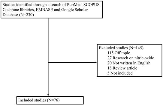
Although various studies have examined the role of ROS in different diseases, the field lacks a published comprehensive literature review comparing the production and role of ROS or free radicals in the development of endometriosis and endometrial cancer. Therefore, we searched five electronic databases—Cochrane Library, EMBASE, Google Scholar, PubMed, and SCOPUS—for studies published between January 1990 and May 2025 using the search terms/strategy ('endometriosis' OR 'endometrial cancer') and ('free radicals' OR 'reactive oxygen species'). The literature search focused on studies published in English and included prospective or retrospective studies on ROS in endometriosis or endometrial cancer performed in humans or animals. Although this study was conducted as a narrative review, we applied a structured screening process solely to ensure the objective and transparent selection of relevant literature for our narrative synthesis, rather than to conduct a formal narrative review. Nevertheless, we applied a structured literature search and screening process to improve transparency and reduce arbitrary article selection. The structured screening process was therefore used to support a comprehensive narrative synthesis and was not intended to fulfill the methodological requirements of a Preferred Reporting Items for Systematic Reviews and Meta-Analyses (PRISMA)-compliant systematic review. A total of 230 articles was initially identified across the five databases. Following a review of titles and abstracts, 154 studies were excluded based on the following predefined criteria: (1) studies focusing on non-endometrial pathologies, (2) articles lacking specific mechanistic data regarding ROS or free radicals, (3) review articles or case reports, (4) unpublished data, gray literature, or non-peer-reviewed materials, and (5) duplicate records. The rationale for selecting the final references was to prioritize studies that provided in-depth mechanistic data regarding specific redox signaling pathways (e.g., PI3K/AKT, NF-κB, ferroptosis, or pyroptosis) rather than purely observational biomarker reports. This approach ensured a robust evidence base for comparing the divergent roles of ROS between the two diseases. Ultimately, 76 articles that met our criteria for thematic relevance and scientific rigor were included for this comprehensive narrative synthesis (Figure 1).
Literature selection process. This diagram summarizes the literature identification and screening process used for this narrative review and is not intended to represent a PRISMA flow diagram.
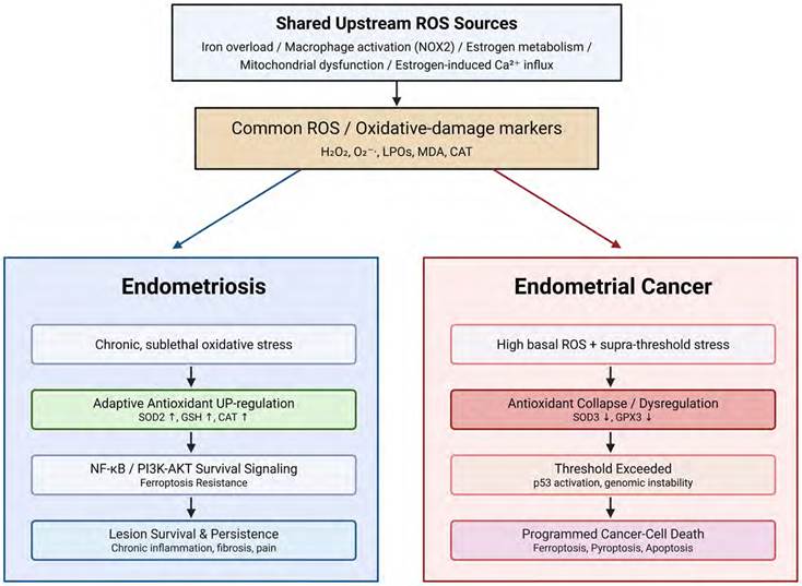
Comparative redox pathway schematic of ROS in endometriosis and endometrial cancer.
3. Discussion
ROS and free radicals are highly reactive molecules produced during cellular metabolic processes. At physiological levels, they contribute to cellular signaling, immune responses, and cell survival. However, excessive accumulation can lead to oxidative stress, resulting in cellular damage and the development of various diseases. Notably, ROS play a central role in the onset and progression of both endometriosis and endometrial cancer, exhibiting contrasting pathophysiological functions depending on the histological nature and microenvironment of each disease. In endometriosis, ROS function within a benign, estrogen-dependent environment, whereas in endometrial cancer, they operate in a malignant setting characterized by genomic instability. This study aims to compare the production and functional roles of ROS and free radicals in endometriosis and endometrial cancer, with the goal of supporting future research on disease pathogenesis and the development of targeted therapeutic strategies. However, the available evidence was not evenly distributed between the two diseases. In this review, fewer studies directly addressed ROS-related mechanisms in endometriosis than in endometrial cancer. Therefore, the comparative interpretation should be understood as a synthesis of currently available mechanistic evidence rather than a quantitatively balanced comparison. In particular, conclusions regarding endometriosis should be interpreted with caution, as several mechanisms remain less extensively characterized than those described in endometrial cancer.
To better conceptualize the opposing roles of ROS, the effects of ROS can be interpreted using a threshold-dependent redox model. At physiological or mildly increased levels, ROS function mainly as signaling molecules that regulate cellular proliferation, inflammatory responses, and adaptive survival pathways. At chronically elevated but sublethal levels, ROS may activate NF-κB, PI3K/AKT, HIF-1α, and related signaling pathways, thereby promoting inflammation, fibrosis, angiogenesis, lesion persistence, and tumor progression. In contrast, when ROS accumulation exceeds the buffering capacity of antioxidant systems such as GSH/GPX4, SOD/CAT, and PRDX, oxidative stress crosses a lethal threshold and induces irreversible cellular injury, including apoptosis, ferroptosis, and pyroptosis. Therefore, the biological effect of ROS is not determined simply by their presence or absence, but by their relative level, duration of exposure, antioxidant reserve, and disease-specific cellular context.
In this framework, endometriotic lesions are generally exposed to chronic, sublethal oxidative stress driven by iron overload, macrophage activation, and estrogen-related metabolism. Rather than immediately inducing cell death, this redox environment may promote inflammatory signaling and lesion survival, particularly when antioxidant defenses are adaptively upregulated. By contrast, endometrial cancer cells often exhibit high basal ROS levels together with altered antioxidant regulation. This makes them vulnerable to therapeutic strategies that further increase ROS beyond a cytotoxic threshold, thereby triggering programmed cell death. Thus, the apparently opposing roles of ROS in endometriosis and endometrial cancer can be explained by a threshold-dependent and context-dependent redox model.
Roles and Mechanisms of Shared ROS Markers in Endometriosis and Endometrial Cancer
| Category & Marker | Endometriosis | Endometrial Cancer | |
|---|---|---|---|
| ROS | H₂O₂ | Induces local oxidative stress; regulated by estrogen to prevent apoptosis; estrogen upregulates GSH → protects cells from H₂O₂-induced apoptosis | Promotes mitochondrial dysfunction and tumor progression; estrogen-induced Ca²⁺ influx increases mitochondrial ROS → EMT, invasion |
| O₂⁻· | Contributes to local inflammation through macrophage activation; generated by NOX2 in activated macrophages | Supports oncogenic signaling and oxidative DNA damage; involved in mitochondrial ROS production and DNA mutation | |
| Oxidative Damage Markers | LPOs/ MDA | Markers of lipid peroxidation and membrane damage; elevated MDA/4-HNE in lesions and serum → fibrosis, pain | Triggers ferroptosis in oxidative conditions; lipid ROS accumulation disrupts membrane integrity → cell death |
| 8-OHdG | Indicates DNA oxidative damage in lesions; ROS attack on guanine → base modification | Promotes genomic instability and tumorigenesis; persistent 8-OHdG lesions cause replication errors | |
| Antioxidant Defense System | GSH | Protects against oxidative cell death; regulated by estrogen to reduce ROS burden | Inhibits ferroptosis under stress; high GSH buffers lipid ROS → blockade of cell death |
| SOD/ SOD3 | Neutralizes O₂⁻·, antioxidant defense; SOD2/MnSOD overexpressed in lesions | SOD3 downregulation → ROS accumulation; loss of SOD3 enhances oxidative stress | |
| Catalase | Reduces H₂O₂; upregulated to compensate for ROS | Present but less effective with tumor burden; insufficient CAT allows H₂O₂ accumulation | |
| HO-1 | Mediates anti-inflammatory macrophage signaling; induced by TGF-β1; protects endometrial cells | Antioxidant defense regulator in the tumor microenvironment; modulates ROS balance and immune escape | |
Abbreviations: 8-OHdG, 8-hydroxy-2'-deoxyguanosine; EMT, epithelial-mesenchymal transition; GSH, glutathione; HO-1, heme oxygenase-1; LPOs, lipid peroxides; MDA, malondialdehyde; MnSOD, manganese superoxide dismutase; NOX2, NADPH oxidase 2; ROS, reactive oxygen species; SOD, superoxide dismutase; TGF-β1; transforming growth factor beta 1; 4-HNE, 4-hydroxynenal; CAT, catalase
Representative ROS-related Regulatory Factors in Endometriosis and Endometrial Cancer
| Disease Context | Regulator Type | Representative Agents / ROS-related Factors | Main Functional Effect |
|---|---|---|---|
| Endometriosis | ROS-attenuating factors | Vitamin C/E, genistein, miR-455, NRF2-related pathways | Reduce oxidative stress and inflammatory signaling; may help attenuate pain and lesion persistence. |
| Endometriosis | ROS-promoting factors | NOX2, TNF, TLRs, MMP-3, P66SHC | Increase oxidative stress, tissue damage, fibrosis, and pain signaling. |
| Endometrial Cancer | ROS-associated tumor progression | Mitochondrial ROS, PKD2/PRDX2/AQP1, S1P, insulin/SFA-related ROS | Promote DNA damage, EMT/angiogenesis, survival signaling, and immune evasion. |
| Endometrial Cancer | ROS-mediated tumor suppression/cell death | Simvastatin, methoxyeugenol, α-terthienylmethanol, sulfasalazine, H₂, ferroptosis/pyroptosis pathways | Increase ROS beyond antioxidant tolerance and induce apoptosis, ferroptosis, or pyroptosis in preclinical models. |
Abbreviations: AQP1, aquaporin-1; AMPK, AMP-activated protein kinase; BCL-2, B-cell lymphoma 2; Ca²⁺, calcium ion; CaBP-28k, calcium-binding protein 28kDa; EC, endometrial cancer; EMT, epithelial-mesenchymal transition; ER, endoplasmic reticulum; ERK, extracellular signal-regulated kinase; EST-1, epithelium-specific ETS transcription factor 1; FHC, ferritin heavy chain; HIF-2α, hypoxia-inducible factor 2 alpha; Hinokitiol, β-thujaplicin; MAPK, mitogen-activated protein kinase; Maackiain, a natural pterocarpan compound; MMP-3, matrix metallopeptidase 3; mTOR, mechanistic target of rapamycin; NADPH, nicotinamide adenine dinucleotide phosphate; NLRP3, NOD-like receptor family pyrin domain containing 3; NOX2, NADPH oxidase 2; NRROS, negative regulator of reactive oxygen species; Nrf2, nuclear factor erythroid 2-related factor 2; P66SHC, 66-kDa SHC adaptor protein; p21, cyclin-dependent kinase inhibitor 1A; p53, tumor protein p53; PGC-1α, peroxisome proliferator-activated receptor gamma coactivator 1-alpha; PKD2, polycystin-2; PRDX2, peroxiredoxin 2; RAS, rat sarcoma virus oncogene family; ROS, reactive oxygen species; S1P, sphingosine-1-phosphate; SFA, saturated fatty acid; Simvastatin, a lipid-lowering statin drug; TLR1/2/4, Toll-like receptor 1/2/4; TNF, tumor necrosis factor; Ucp2, uncoupling protein 2; UPBEAT1, upstream binding transcription factor 1; Vitamin C/E, ascorbic acid / α-tocopherol; α-Terthienylmethanol, a thiophene-based redox-active compound.
3.1. ROS, free radicals, and related biomarkers specifically expressed in endometrial cancer
In previous studies conducted by our department, we identified a number of ROS and free radicals in endometriosis, including H₂O₂, O₂⁻·, ·OH, lipid peroxides (LPOs), malondialdehyde (MDA), 4-hydroxynonenal (4-HNE), GSH, SOD2, GPx, CAT, NAD(P)H:quinone oxidoreductase 1 (NQO1), thioredoxin (TRX), and peroxiredoxins (PRDX). In endometrial cancer, the ROS and free radicals detected included H₂O₂, O₂⁻·, LPOs, MDA, SOD3, CAT, PRDX2, PKD2, and heme oxygenase-1 (HO-1). Among these, H₂O₂, O₂⁻·, LPOs, MDA, and CAT were commonly expressed in both endometriosis and endometrial cancer. However, SOD3, PKD2, PRDX2, and HO-1 were exclusively observed in endometrial cancer and are thought to contribute to its pathogenesis. In endometrial cancer, ROS do not merely act as metabolic byproducts but also function as factors regulating apoptosis, immune responses, tumor suppression, or progression. In particular, various ROS-related factors are specifically observed in endometrial cancer, which is an important characteristic demonstrating the pathophysiological differences from endometriosis [29,30].
SOD3 is specifically downregulated in endometrial cancer, leading to the accumulation of excess O₂⁻·, which in turn promotes DNA damage and tumor invasiveness. While overexpression of SOD2 has been observed in endometriosis, suppression of SOD3 has not been reported, suggesting that SOD3 may serve as a cancer-specific antioxidant regulator in endometrial malignancies.
Both PRDX2 and PKD2 play key roles in oxidative stress regulation and cancer cell survival in endometrial cancer. PRDX2 is overexpressed and protects cancer cells from oxidative damage by scavenging excess ROS, thereby contributing to tumor survival and potential therapeutic resistance. PKD2, on the other hand, activates pro-survival pathways such as the phosphoinositide 3-kinase (PI3K)/protein kinase B (AKT) cascade in response to ROS-induced calcium signaling. Overexpression of PKD2 has been associated with altered expression of microRNAs, including miR-195-3p, miR-20a and miR-134, suggesting the possibility of indirect regulation via miRNA pathways. These expression patterns have not been clearly reported in endometriosis.
Collectively, these findings indicate that certain ROS-related molecules exhibit distinct expression patterns and functional roles specific to endometrial cancer, underscoring their potential as therapeutic targets.
3.2. The Dual Role of ROS-Mediated Cell Death: Pathogenesis in Endometriosis vs. Tumor Suppression in Endometrial Cancer
In endometriosis, ROS contribute to key pathophysiological mechanisms such as chronic inflammation, fibrosis, pain, and infertility. Representative ROS include H₂O₂, O₂⁻·,LPOs and MDA, which are primarily produced by activated peritoneal macrophages and iron overload. These ROS disrupt the redox balance of the peritoneal microenvironment, leading to tissue damage. In contrast, the same ROS can inhibit tumor growth in endometrial cancer by promoting regulated forms of cancer cell death.
Ferroptosis is a form of regulated cell death characterized by the iron-dependent accumulation of lethal LPOs. The canonical upstream regulatory cascade of ferroptosis primarily hinges on the System Xc⁻/GSH/GPX4 axis [31]. Under physiological conditions, GPX4 utilizes GSH to reduce toxic lipid hydroperoxides into non-toxic lipid alcohols. However, when GPX4 is inactivated or GSH is depleted, this antioxidant defense fails. Consequently, iron-dependent enzymes (such as 15-lipoxygenase) and intracellular loosely-bound iron synergistically catalyze the highly selective oxidation of polyunsaturated fatty acids (e.g., arachidonoyl-phosphatidylethanolamines) and drive subsequent Fenton-like cleavage reactions [32]. This dual enzymatic and free-radical cascade triggers massive accumulation of electrophilic lipid oxidation products and subsequent membrane rupture.
The pathophysiological implications of this cascade differ significantly between endometriosis and endometrial cancer. In endometriosis, retrograde menstruation repeatedly exposes the peritoneal cavity to an iron-overloaded microenvironment [29]. While this iron toxicity initially drives early lesion clearance, surviving ectopic endometrial cells adaptively upregulate antioxidant defenses to establish ferroptosis resistance, thereby sustaining lesion survival and chronic inflammation [33]. Furthermore, recent evidence highlights that ectopic cells actively fortify this resistance by orchestrating survival pathways, such as the PDLIM3/Gli1/Hedgehog axis, to suppress iron-dependent LPO accumulation [34]. Conversely, in endometrial cancer, malignant cells depend highly on the GPX4/SLC7A11 pathway to buffer their elevated metabolic ROS. Consequently, targeted therapeutic agents that inhibit the RAS/mitogen-activated protein kinase (MAPK) pathway or activate the ROS/ AMP-activated protein kinase (AMPK)/mechanistic target of rapamycin (mTOR) axis can actively dismantle this antioxidant defense to induce lipid peroxidation and selectively trigger ferroptosis in cancer cells [35]. Therefore, while overcoming ferroptosis resistance is critical for clearing benign endometriotic lesions, the active induction of ferroptosis may represent a promising investigational strategy for targeting malignant endometrial cancer cells.
Pyroptosis is a highly inflammatory form of programmed cell death driven by the inflammasome cascade. The canonical activation of the NOD-, LRR- and pyrin domain-containing protein 3 (NLRP3) inflammasome involves a tightly regulated sequence: First, ROS-dependent activation of the NF-κB pathway drives the transcriptional induction of NLRP3 and pro-IL-1β. Second, a subsequent activation signal—often triggered by severe oxidative stress and intracellular potassium efflux—promotes the oligomerization of NLRP3. This oligomer recruits the adaptor protein, apoptosis-associated speck-like protein containing a CARD, to assemble a macromolecular inflammasome complex, which subsequently recruits and cleaves pro-caspase-1 into its active form. Finally, active caspase-1 cleaves gasdermin-D (GSDMD) to release its N-terminal domain to form pores in the plasma membrane and thereby execute rapid cell lysis [36-38].
The therapeutic implications of this ROS-driven pyroptotic cascade contrast sharply between endometrial cancer and endometriosis. In endometrial cancer, experimental ROS induction has been investigated as a potential way to activate this NLRP3/caspase-1/GSDMD axis. Activating pyroptosis may contribute to malignant cell death and modulation of tumor immune evasion by altering the tumor microenvironment. In endometriosis, in contrast, chronic activation of the NLRP3 inflammasome has been directly linked to lesion progression and angiogenesis. Within this inherently inflammatory microenvironment, continuous ROS-induced pyroptosis exacerbates ectopic lesions and sustains chronic inflammation [39,40]. Therefore, while triggering pyroptosis may represent a potential anti-tumor mechanism in endometrial cancer, targeted inhibition of the NLRP3 inflammasome cascade remains a rational and beneficial therapeutic strategy for endometriosis.
3.3. Crosstalk Between PI3K/AKT and NF-κB Signaling Pathways
To fully delineate the pathophysiological impact of ROS, it is essential to integrate the PI3K/AKT and NF-κB signaling networks. ROS accumulation triggers intracellular calcium flux, which activates protein kinase D2 to phosphorylate and drives the PI3K/AKT survival cascade [41]. Concurrently, ROS serves as a potent upstream activator of the NF-κB transcription factor, which translocates to the nucleus to drive the expression of pro-inflammatory and anti-apoptotic genes [42]. The synergistic crosstalk between these two pathways dictates divergent cell fates in endometriosis and endometrial cancer. In endometriosis, this integration establishes a vicious cycle in which PI3K/AKT signaling promotes the aberrant survival and proliferation of ectopic cells [43], while NF-κB sustains a chronic inflammatory microenvironment [44]. Together, they fortify the resistance of endometriotic lesions to physiological clearance. Conversely, in endometrial cancer, although these integrated pathways initially facilitate tumor progression and immune evasion, this network is highly sensitive to redox imbalances. Therapeutic agents that increase ROS levels may overwhelm adaptive PI3K/AKT and NF-κB survival signals in experimental settings, thereby shifting the cellular balance toward apoptotic, ferroptotic, or pyroptotic cell death. Thus, the intersection of calcium-dependent PI3K/AKT and ROS-driven NF-κB cascades represents a critical regulatory hub in both diseases.
3.4. Opposing Directional Regulation of Antioxidant Systems
Antioxidant defense systems tend to be upregulated in endometriotic lesions, serving to protect cells from ROS and sustain lesion viability. Notably, the expression of antioxidant enzymes such as SOD2, GSH, and CAT is significantly upregulated in endometriotic tissue, enabling lesion survival under oxidative stress conditions [5,8]. In addition, miR-455 plays a protective role by suppressing fatty acid-binding protein 4 (FABP4), thereby reducing H₂O₂-induced cell death and limiting ROS accumulation [45]. Estrogen also promotes GSH biosynthesis and increases CAT activity, effectively inhibiting ROS-mediated cell death—a mechanism closely linked to the estrogen-dependent survival characteristic of endometriosis [46,47].
In contrast, endometrial cancer is characterized by the collapse or downregulation of antioxidant systems. The expression of SOD3 is significantly reduced, impairing the clearance of extracellular O2-· and accelerating ROS accumulation within the tumor microenvironment. Additionally, antioxidant enzymes such as glutathione peroxidase 3 (GPX3), PRDX2, and CAT are either functionally lost or downregulated, rendering cancer cells more susceptible to oxidative stress and promoting cell death pathways such as ferroptosis and apoptosis [35,48,49].
These contrasting patterns indicate that antioxidant regulation differs according to disease context. In endometriosis, adaptive antioxidant responses may support lesion persistence under chronic oxidative stress, whereas in endometrial cancer, altered antioxidant regulation may influence susceptibility to ROS-mediated cell death. This disease-specific directionality provides a mechanistic basis for the therapeutic discussion below.
3.5. Differences in the Immune Microenvironment
Endometriosis and endometrial cancer exhibit distinct immune microenvironments that are closely associated with redox balance. In endometriosis, peritoneal macrophages express HO-1, contributing to an immunotolerant environment that supports the long-term survival of endometriotic lesions. The transforming growth factor beta 1 (TGF-β1)-HO-1 signaling axis also enables these lesions to evade ROS-mediated cellular injury. In contrast, endometrial cancer is characterized by an immunosuppressive tumor microenvironment driven by high metabolic activity and altered redox balance. Through the Warburg effect, highly proliferative cancer cells rely on aerobic glycolysis, which leads to lactate accumulation and the formation of an acidic microenvironment. This acidic pH interacts with elevated ROS levels and disrupts immune surveillance. Together, acidic pH and elevated ROS levels promote the differentiation of regulatory T cells (Tregs) and M2 macrophages. Acidic pH also impairs the cytolytic activity of CD8+ T cells. In addition, suppression of the NLRP3 inflammasome and a decrease in the ceramide-to-sphingosine-1-phosphate (S1P) ratio contribute to the development of this immunosuppressive milieu. Consequently, the interaction between acidic pH and ROS accumulation creates a vicious cycle that facilitates immune evasion and tumor progression. This mechanism is fundamentally distinct from the iron-driven oxidative stress observed in endometriotic lesions.
3.6. Justification for Disease-Specific Therapeutic Strategies
Therapeutic approaches targeting ROS should account for the disease-specific oxidative environment, but their clinical implications should be interpreted according to the level of supporting evidence. In endometriosis, antioxidant strategies have been investigated as a way to attenuate ROS-associated inflammation and pain. Vitamins C and E, genistein, nanoceria, and miR-455-related mechanisms have been discussed in experimental or clinical contexts, although the strength of evidence differs across interventions [45,50,52,53]. In endometrial cancer, ROS-inducing approaches have been explored mainly in preclinical studies as a means of increasing oxidative stress beyond the tolerance of malignant cells. Agents such as hinokitiol, simvastatin, α-terthienylmethanol, and molecular hydrogen have been investigated for their ability to modulate ROS-related cell death pathways [35,48,51]. Therefore, antioxidant and pro-oxidant strategies should be regarded as disease-context-dependent concepts requiring further validation rather than uniformly established therapeutic approaches.
Furthermore, it is important to recognize the context-dependent dual nature of many anti-inflammatory and antioxidant compounds, such as vitamin C and certain polyphenols. In the benign, chronic inflammatory microenvironment of endometriosis, these compounds may primarily function as cytoprotective agents by scavenging ROS and mitigating tissue damage and pain. However, in the context of endometrial cancer, the same compounds may act as pro-oxidants under specific conditions. At high pharmacological concentrations or in the presence of transition metals, these compounds may undergo auto-oxidation and generate H₂O₂ through Fenton-like reactions. Because endometrial cancer cells may exhibit altered antioxidant defense mechanisms, this increase in radical production could exceed their oxidative threshold and contribute to apoptosis or ferroptosis [55,56]. Thus, the ability of these compounds to act protectively or promote radical production is dictated by their dosage and the specific redox status of the surrounding cellular microenvironment.
3.7. Relationship Between ROS Mechanisms and Molecular Features of Endometrial Cancer
To improve the translational relevance of this review, ROS-related mechanisms should be interpreted in the context of the molecular features of endometrial cancer defined by The Cancer Genome Atlas (TCGA). As described above, TCGA classifies endometrial cancer into four molecular subtypes: POLE-ultramutated, microsatellite instability-high/mismatch repair-deficient, copy-number low, and copy-number high/p53-abnormal [13,14].
However, the available evidence was insufficient to directly assign specific ROS-related mechanisms or ROS thresholds to individual TCGA molecular subtypes. Therefore, we did not draw subtype-specific conclusions in the present review.
Nevertheless, several ROS-related mechanisms discussed here overlap with molecular features that are relevant to endometrial cancer biology. These include genomic instability, p53-related cell-cycle regulation, PTEN/PI3K/AKT signaling, NF-κB-mediated inflammatory signaling, and altered antioxidant regulation [11-14]. Excessive ROS accumulation can induce DNA damage, genomic instability, and cell-cycle dysregulation. In addition, ROS-related pathways, including PI3K/AKT and NF-κB signaling, may influence cancer cell survival, inflammation, immune evasion, and therapeutic response [17-19,41,42]. Altered antioxidant regulators, such as SOD3, PRDX2, PKD2, GSH, and GPX4, may also contribute to redox balance. This balance may determine whether cancer cells survive or undergo programmed cell death [17,18,31,35,57,58].
Thus, rather than establishing a definitive subtype-specific ROS model, this review proposes that the TCGA molecular classification may serve as a useful framework for future studies. In particular, it may help clarify how ROS-related mechanisms differ among molecularly defined subgroups of endometrial cancer. Further subtype-stratified studies are needed to determine whether ROS levels, antioxidant capacity, and sensitivity to ROS-inducing therapies vary according to TCGA subtype.
3.8. Opposing Roles of Shared Markers in Endometriosis and Endometrial Cancer
Several ROS-related biomarkers play contrasting roles in endometriosis and endometrial cancer. For instance, GSH acts as a cytoprotective agent in endometriosis, whereas in endometrial cancer, it suppresses ferroptosis and contributes to therapeutic resistance. Similarly, lipid peroxidation products such as MDA and 4-HNE promote fibrosis and pain in endometriosis but serve as inducers of ferroptosis and cell death in endometrial cancer. SOD2 exerts antioxidant effects in endometriosis, whereas in endometrial cancer, decreased SOD3 expression leads to ROS accumulation and increased sensitivity to cell death.
H₂O₂
In endometriosis, H₂O₂ functions primarily as a pro-inflammatory mediator and lesion-sustaining factor. As endometrial tissue migrates outside the uterus, it triggers inflammatory responses; peritoneal macrophages and lesion cells produce large amounts of ROS, including H₂O₂, which induces oxidative stress and damages cellular components such as membranes, proteins, and DNA. Endometriotic lesions show an imbalance in antioxidant defenses: SOD is overactive, converting O₂⁻·into H₂O₂, but CAT and GPx, which are responsible for degrading H₂O₂, are downregulated [5]. This imbalance facilitates intracellular accumulation of H₂O₂, which activates the NLRP3 inflammasome, induces pyroptosis, and contributes to chronic inflammation, invasion, and pain. In contrast, in endometrial cancer, H₂O₂ promotes genomic instability and carcinogenesis by inducing oxidative DNA damage, p53 mutations, and cell-cycle dysregulation. Interestingly, H₂O₂ has also emerged as a therapeutic target: when ROS levels exceed the antioxidant capacity of cancer cells, it can trigger apoptosis or ferroptosis. Thus, H₂O₂-inducing approaches are being investigated in preclinical studies as a potential anticancer strategy that requires further validation.
GSH
In endometriosis, GSH protects cells against oxidative stress. Exposure of endometrial stromal cells to H₂O₂ in the presence of estrogen (17β-estradiol) elevates GSH levels, inhibiting apoptosis and stabilizing mitochondrial membrane potential and thereby promoting lesion survival. GSH thus facilitates chronic persistence of lesions and is being investigated as a therapeutic target, with studies exploring GSH inhibition to reduce lesion viability. In endometrial cancer, GSH is crucial in regulating oxidative stress, particularly in evading ferroptosis and promoting drug resistance. It cooperates with GPX4 to detoxify LPOs, enabling cancer cell survival and metabolic adaptation. High GSH levels and increased cystine/glutamate transporter (xCT) expression have been observed in uterine serous carcinoma (USC), contributing to cisplatin resistance. xCT inhibition by sulfasalazine depletes GSH, causing ROS accumulation and enhancing cell death, underscoring the key role of GSH in cancer cell survival and drug resistance. These findings suggest that GSH metabolism may serve as a promising therapeutic target in endometrial cancer [57,58].
SOD2 versus SOD3
Antioxidant enzymes from the SOD family exhibit disease-specific effects. In endometriosis, mitochondrial SOD2 expression is upregulated, converting excess intracellular O₂⁻· to H₂O₂ and alleviating oxidative stress. This process suppresses cell death and is enhanced by estrogen, which promotes mitochondrial stability and lesion persistence by inhibiting caspase-3 activation. In contrast, SOD3, an extracellular isoform, is downregulated in endometrial cancer, leading to ROS accumulation in the tumor microenvironment. This increases cancer cell susceptibility to oxidative cell death processes such as ferroptosis and may enhance sensitivity to anticancer therapies. Thus, whereas SOD2 supports lesion survival in endometriosis, an SOD3 deficiency facilitates tumor cell death in endometrial cancer, highlighting how the same antioxidant system can yield opposite outcomes depending on disease context.
LPOs and MDA
LPOs and MDA—initial products and secondary byproduct, respectively, of lipid peroxidation—are shared markers of oxidative stress but play different pathophysiological roles in the two diseases. In endometriosis, chronic inflammation and ROS accumulation promote lipid peroxidation, oxidizing membrane polyunsaturated fatty acids and generating MDA. MDA exacerbates tissue damage and inflammation and is associated with pain. Iron-rich and ROS-dense peritoneal environments further accelerate lipid peroxidation, supporting lesion invasiveness and chronic pain. In endometrial cancer, MDA is not merely a marker of damage but is also an indicator of ferroptosis activation. Accumulation of LPOs and MDA within cancer cells reflects engagement of ferroptotic pathways, making MDA a potential predictive biomarker of anticancer responsiveness. Thus, while LPOs and MDA contribute to pathology and pain in endometriosis, they promote therapeutic cell death in endometrial cancer, underscoring the contextual duality of these shared oxidative markers.
4. Clinical Implications and Therapeutic Strategies Targeting the Redox Balance
Given the contrasting roles of ROS in endometriosis and endometrial cancer, redox-targeted therapeutic strategies may need to be tailored to the disease context. However, these therapeutic implications should be interpreted cautiously, because the level of evidence differs substantially between preclinical models and clinically validated interventions. Antioxidant approaches in endometriosis are supported by experimental studies and some clinical evidence, whereas pro-oxidant strategies in endometrial cancer remain largely preclinical and require further validation before clinical application (Table 3 and Figure 3).
Summary of Key Representative Studies on ROS Modulation
| Disease Category | Author (Year) [Ref] | Study Design & Model | Target Agent / Factor | Key ROS-related Findings & Outcomes |
|---|---|---|---|---|
| Endometriosis | Ngô et al. (2009) [5] | In vitro & In vivo - Primary cells, Nude mice | ROS, N-acetyl-cysteine (NAC) | Endometriotic cells show a pro-proliferative phenotype driven by high ROS-induced ERK1/2 activation; NAC suppresses lesion development. |
| Xie et al. (2017) [6] | Clinical & In vitro (Cell) - Human tissues, Primary cells | ROS (H₂O₂), ARID1A, DNMT1 | ROS exposure decreases the tumor suppressor ARID1A by inducing hyper-methylation of its promoter via DNMT1 upregulation. | |
| Sutrisno et al. (2022) [7] | In vivo (Animal) - Mice model | Genistein | Genistein acts as an antioxidant by significantly increasing SOD and GPx levels in peritoneal fluid, preventing oxidative stress. | |
| Li et al. (2021) [33] | In vitro (Cell) & In vivo (Animal) - Primary cells, C57BL/6 mice | Erastin, ferroportin | Erastin induces ferroptosis in ectopic stromal cells by triggering iron-dependent ROS accumulation, reducing lesion growth in vivo. | |
| Tang et al. (2019) [45] | In vitro (Cell) - Immortalized HESCs | miR-455, FABP4, ROS (H₂O₂) | miR-455 protects cells from H₂O₂-induced oxidative stress by downregulating FABP4 and thereby lowering ROS and lipid oxidation. | |
| Andrade et al. (2013) [47] | In vitro (Cell) - Primary stromal cells | 17β-Estradiol, H₂O₂ | 17β-Estradiol combined with steady-state H₂O₂ exerts an antiapoptotic effect, enhancing cell proliferation and ERK phosphorylation. | |
| Santanam et al. (2012) [50] | Clinical - Human patients | Antioxidant vitamins (Vit E & C) | Antioxidant supplementation significantly reduces chronic pelvic pain and decreases inflammatory markers in peritoneal fluid. | |
| Endometrial Cancer | Zhou et al. (2022) [35] | In vitro (Cell) - Ishikawa cells | Simvastatin | Suppresses proliferation and induces ferroptosis by increasing ROS, MDA, and iron levels while reducing GSH via RAS/MAPK suppression. |
| Lee et al. (2015) [48] | In vitro (Cell) - Hec1A and Ishikawa cells | α-Terthienylmethanol, NADPH oxidase | Induces apoptosis by promoting intracellular ROS accumulation and depleting GSH, mediated partially through NADPH oxidase activation. | |
| Costa et al. (2021) [51] | In vitro (Cell) - Human endometrial cancer cells | Methoxyeugenol | Induces an increase in ROS, which regulates the p53/p21 pathway and effectively suppresses human endometrial cancer cell proliferation. | |
| Sendo et al. (2022) [57] | In vitro (Cell) & In vivo (Animal) - Carcinoma cells, SCID mice | Sulfasalazine, GSH, cisplatin | Enhances cisplatin-induced apoptosis by depleting intracellular GSH and causing massive ROS accumulation. |
Abbreviations: ARID1A, AT-rich interactive domain-containing protein 1A; DNMT1, DNA methyltransferase 1; ERK, extracellular signal-regulated kinase; FABP4, fatty acid binding protein 4;GPx, glutathione peroxidase; GSH, glutathione; H₂O₂, hydrogen peroxide; MAPK, mitogen-activated protein kinase; MDA, malondialdehyde; NAC, N-acetyl-cysteine; ROS, reactive oxygen species; SCID, severe combined immunodeficiency; SOD, superoxide dismutase
Divergent therapeutic strategies targeting ROS in endometriosis and endometrial cancer. Shared ROS sources and markers converge on a common mechanistic core—the System Xc⁻ (SLC7A11)/GSH/GPX4 axis—whose redox threshold sets the divergent outcome. In endometriosis (top right), ROS accumulation drives inflammation and lesion survival, and antioxidant strategies aim to hold ROS below the inflammatory threshold. Conversely, in endometrial cancer (bottom right), massive ROS induction may act as a potential pro-oxidant stimulus in preclinical models. Pro-oxidant agents overwhelm the collapsed antioxidant defenses of tumor cells, driving lipid peroxidation and resulting in programmed cell death such as ferroptosis and pyroptosis.
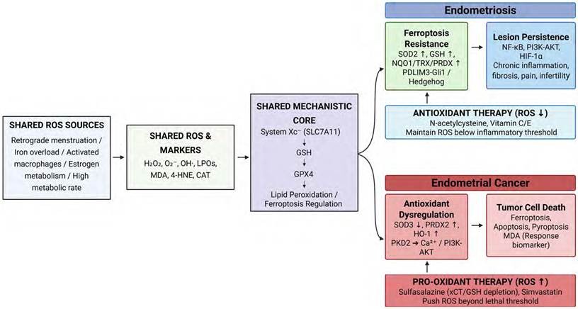
Table 3 summarizes representative experimental and clinical studies that directly modulated ROS in endometriosis or endometrial cancer, allowing their divergent roles to be compared side by side. In the endometriosis studies, interventions that lowered ROS or reinforced antioxidant defenses — such as N-acetyl-cysteine, genistein, and vitamins C/E — attenuated lesion development, restored SOD/GPx activity, and reduced pelvic pain and inflammatory markers. In the endometrial cancer studies, the opposite therapeutic logic applied: agents such as simvastatin, α-terthienylmethanol, methoxyeugenol, and sulfasalazine suppressed tumor cell proliferation by increasing ROS and depleting GSH, thereby driving apoptosis or ferroptosis. Taken together, these studies demonstrate that the same molecular currency — ROS and the GSH/SOD antioxidant axis — is exploited in opposite directions across the two diseases, directly supporting our central conclusion that redox-targeted therapy must be disease-context-specific: antioxidant (ROS-lowering) in endometriosis and pro-oxidant (ROS-raising) in endometrial cancer. It should be noted, however, that most endometrial cancer entries derive from in vitro or animal models, underscoring that these pro-oxidant strategies remain investigational.
4.1. Antioxidant Therapies in Endometriosis
In endometriosis, the primary therapeutic goal is to attenuate the ROS-driven cycle of inflammation and lesion survival. Preclinical studies suggest that antioxidants may mitigate disease progression by reducing ROS-dependent inflammatory and proliferative signaling. For instance, N-acetyl-cysteine has been shown to suppress lesion development by inhibiting ROS-induced ERK1/2 activation in vivo [5], while genistein restored peritoneal antioxidant enzyme activity, including SOD and GPx, in an experimental model [7].
Clinically, antioxidant supplementation has shown potential symptom-relieving effects in endometriosis. Clinical studies involving vitamins C and E reported reductions in chronic pelvic pain and inflammatory markers in peritoneal fluid [50], and recent meta-analyses have suggested beneficial effects of antioxidant supplementation on pain and oxidative stress markers, although the magnitude of effect varied across outcomes, antioxidant regimens, and included studies [52,53]. However, these clinical findings should be interpreted cautiously because antioxidant regimens, patient populations, disease stages, and outcome measures vary across studies. Therefore, antioxidant therapy should be considered a promising adjunctive strategy rather than a clinically established disease-modifying treatment.
4.2. Pro-oxidant and ROS-Inducing Therapies in Endometrial Cancer
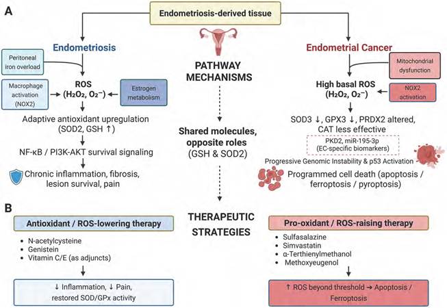
In endometrial cancer, pro-oxidant strategies are based on the concept that malignant cells with high basal ROS levels may be vulnerable to additional oxidative stress. However, most evidence for ROS-inducing therapies in endometrial cancer remains preclinical. Agents such as simvastatin, α-terthienylmethanol, methoxyeugenol, and sulfasalazine have been reported to increase ROS accumulation, deplete antioxidant defenses, or promote apoptosis or ferroptosis in cell-based or animal models [35,48,51,57]. These findings provide a mechanistic rationale for further investigation of ROS-inducing approaches, particularly in relation to ferroptosis, apoptosis, pyroptosis, and chemoresistance. Nevertheless, these strategies have not yet been sufficiently validated as established clinical therapies for endometrial cancer. Differences in experimental models, ROS detection methods, drug concentrations, and tumor molecular context limit direct translation to patient care. Therefore, pro-oxidant therapy should currently be regarded as an investigational approach rather than a clinically established treatment strategy. To synthesize these mechanistic and therapeutic insights, a comprehensive summary of the context-dependent roles of ROS in both diseases is provided in Figure 4.
Context-dependent roles of ROS and targeted therapeutic strategies in endometriosis and endometrial cancer. (A) In endometriosis, sublethal ROS drive adaptive antioxidant upregulation (SOD2, GSH), sustaining survival signaling and lesion persistence. Conversely, in endometrial cancer, supra-threshold ROS cause antioxidant collapse and p53 activation, triggering programmed cell death. Notably, shared mediators (GSH, SOD2) exert opposite roles—promoting survival in endometriosis but chemoresistance in cancer. (B) Consequently, antioxidant therapies lower ROS to mitigate endometriosis, whereas pro-oxidant therapies raise ROS beyond the lethal threshold to induce cancer cell death, underscoring a disease-specific redox model.
5. Limitations
This study compared the roles of ROS and free radicals in the distinct disease contexts of endometriosis and endometrial cancer, evaluating their pathophysiological differences and therapeutic potential. However, several limitations should be noted. First, this review integrated heterogeneous studies conducted under varying experimental conditions. The cell lines, animal models, tissue samples, and ROS detection methods differed across studies, making direct comparison challenging and requiring caution in generalizing results. Second, although more included studies addressed ROS-related mechanisms in endometrial cancer than in endometriosis, detailed mechanistic understanding of specific ROS-induced cell death pathways in endometrial cancer, particularly ferroptosis and pyroptosis, remains incomplete. Quantitative analyses and in vivo validation studies are still limited. Third, ROS are context-dependent molecules whose effects cannot be understood solely based on their presence. Their functions vary drastically depending on concentration, exposure duration, and the metabolic state of the cell. However, this review did not fully account for the temporal dynamics or stage-specific changes in ROS activity, which may have limited interpretability of ROS thresholds and functional transitions. Fourth, as this study was reclassified and conducted as a narrative review rather than a strict systematic review following PRISMA guidelines, there are inherent methodological limitations. Although a comprehensive literature search was performed across multiple major databases, the selection process may be subject to selection bias. Furthermore, relevant studies not indexed in the selected databases, unpublished data, and/or articles published in languages other than English may have been inadvertently omitted. Fifth, the evidence base was not equally distributed between the two disease entities. Fewer included studies directly investigated ROS-related mechanisms in endometriosis than in endometrial cancer, which may have affected the balance and depth of the comparative analysis. As a result, the endometrial cancer sections may provide greater mechanistic specificity, whereas some conclusions regarding endometriosis should be interpreted as hypothesis-generating and requiring further validation. Sixth, the level of evidence differed considerably across therapeutic strategies. Many ROS-targeted interventions, particularly pro-oxidant strategies for endometrial cancer, were supported primarily by cell-based or animal studies rather than clinically validated trials. Even in endometriosis, where antioxidant supplementation has been evaluated in clinical studies, differences in antioxidant type, dose, treatment duration, patient population, disease severity, and outcome measures make it difficult to draw definitive therapeutic conclusions. In addition, potentially conflicting effects of antioxidants and pro-oxidants should be considered, as the same compound may exert different effects depending on dose, redox status, and disease context. Seventh, translating these redox-modulating strategies into clinical practice faces several significant hurdles. A primary concern is treatment specificity and the potential for systemic toxicity. Because ROS act as essential signaling molecules for normal physiological functions and immune surveillance in healthy tissues, the systemic administration of potent pro-oxidant or non-specific antioxidant agents may induce severe off-target effects. Furthermore, patient heterogeneity significantly complicates the establishment of standardized therapeutic regimens. Variations in baseline systemic oxidative stress, diverse genetic backgrounds, and distinct molecular subtypes (such as the TCGA molecular classifications in endometrial cancer) suggest that a uniform redox intervention may yield unpredictable clinical outcomes. Therefore, successful clinical translation will likely depend on the development of targeted delivery systems and robust predictive biomarkers to stratify patients based on their individual redox vulnerabilities.
6. Conclusions
Although ROS are commonly produced in both endometriosis and endometrial cancer, which share the characteristic of abnormal proliferation of endometrial-derived tissue, they exhibit markedly contrasting pathophysiological roles. In endometriosis, ROS are primarily generated through peritoneal iron overload, macrophage activation, and estrogen metabolism. These ROS contribute to chronic inflammation, fibrosis, lesion survival, and pain. Endometriotic lesions adapt to this oxidative stress environment by upregulating antioxidant systems and evolving mechanisms for evading ROS-induced cell death. In contrast, in endometrial cancer, ROS accumulation results from mechanisms such as mitochondrial dysfunction, NOX2 activation, and estrogen-induced Ca2+ influx. These ROS initially mediate oncogenic processes, including DNA damage and tumor progression. However, once ROS levels surpass a certain threshold, they overwhelm the adaptive PI3K/AKT and NF-κB signaling networks, triggering programmed cell death pathways such as apoptosis, ferroptosis, and pyroptosis. Notably, ROS-regulating molecules such as SOD3, miR-195-3p, and protein kinase D2 are uniquely observed in endometrial cancer and show promise as diagnostic or therapeutic biomarkers. Moreover, antioxidant molecules like GSH and SOD2—which function to suppress ROS accumulation and promote lesion survival in endometriosis—paradoxically contribute to therapeutic resistance in endometrial cancer by inhibiting ferroptosis. This underscores the context-dependent and even opposing functions that the same molecules can exert in different disease environments.
Therefore, ROS should not be viewed merely as pathological byproducts, but should rather be redefined as dual-function regulators that can shift between survival and death signals depending on tissue context and microenvironment. This paradigm may inform disease-specific, ROS-targeted therapeutic strategies: in endometriosis, antioxidant approaches aimed at suppressing excessive ROS may help alleviate inflammation and pain, whereas in endometrial cancer, pro-oxidant strategies designed to trigger cancer cell death remain promising but require further clinical validation. Importantly, the dual role of antioxidants such as vitamin C further illustrates that redox-targeted interventions cannot be categorized simply as protective or harmful; rather, their effects depend on dose, redox status, and disease-specific microenvironment. These findings emphasize the need for precision medicine approaches that tailor treatments based on the distinct redox vulnerabilities of each disease. Future efforts to map ROS-centered molecular networks and validate redox-targeted interventions in clinically relevant models and patient cohorts may help refine therapeutic strategies for endometrial disorders.
Conceptualization, J.L., S.G.Y. and D.C.P.; methodology, J.L., J.M.L., H.O.K, M.K.S., S.S.K., T.I.O., J.Y.; validation, J.L., J.M.L., H.O.K, S.S.K., T.I.O., J.Y.; formal analysis, J.M.L., H.O.K, S.S.K., T.I.O., J.Y.; investigation, S.G.Y.; resources, S.G.Y. and D.C.P.; data curation, J.M.L., H.O.K, S.S.K., T.I.O., J.Y.; writing—original draft preparation, J.L., S.G.Y. and D.C.P.; writing—review and editing, S.G.Y. and D.C.P.; visualization, J.L. and S.G.Y.; supervision, D.C.P.; project administration, S.G.Y.; funding acquisition, S.G.Y. and S.S.K. All authors have read and agreed to the published version of the manuscript.
Acknowledgements
Funding
This work was supported by the ministry of Education of the Republic of Korea and the National Research Foundation of Korea (NRF) grant funded by the Korean government (NRF-2018R1A6A1A03025124) (NRF- 2022R1A2C1091779) (NRF-2025S1A5C3A03022311). This research was supported by a grant of National Bio Bigdata Project, funded by four ministries (Ministry of Health and Welfare, Ministry of Science and ICT, Ministry of Trade, Industry and Re-sources, and Korea Disease Control and Prevention Agency) of Korea (grant number: RS-2024-00509118). The funders had no role in study design, data collection, data analysis, data interpretation, or writing of the report.
Competing Interests
The authors have declared that no competing interest exists.
References
1. Dunselman GAJ, Vermeulen N, Becker C, Calhaz-Jorge C, D'Hooghe T, De Bie B. et al. ESHRE guideline: management of women with endometriosis. Hum Reprod. 2014;29:400-12
2. Kim H, Cho SH. Diagnosis and treatment of endometriosis. J Korean Med Assoc. 2019;62:513-8
3. Dinulescu DM, Ince TA, Quade BJ, Shafer SA, Crowley D, Jacks T. Role of K-ras and Pten in the development of mouse models of endometriosis and endometrioid ovarian cancer. Nat Med. 2005;11:63-70
4. Hevir N, Ribič-Pucelj M, Lanišnik Rižner T. Disturbed balance between phase I and II metabolizing enzymes in ovarian endometriosis: a source of excessive hydroxy-estrogens and ROS? Mol Cell Endocrinol. 2013;367:74-84
5. Ngô C, Chéreau C, Nicco C, Weill B, Chapron C, Batteux F. Reactive oxygen species controls endometriosis progression. Am J Pathol. 2009;175:225-34
6. Xie H, Chen P, Huang HW, Liu LP, Zhao F. Reactive oxygen species downregulate ARID1A expression via its promoter methylation during the pathogenesis of endometriosis. Eur Rev Med Pharmacol Sci. 2017;21:4509-15
7. Sutrisno S, Miryani I, Made Dwijayasa P, Rini Suprobo N, Wayan Arsana Wiyasa I. Genistein administration increases the level of superoxide dismutase and glutathione peroxidase in the endometriosis mice model: an experimental study. Int J Reprod Biomed. 2022;20:873-82
8. Chen C, Zhou Y, Hu C, Wang Y, Yan Z, Li Z. et al. Mitochondria and oxidative stress in ovarian endometriosis. Free Radic Biol Med. 2019;136:22-34
9. Oaknin A, Bosse TJ, Creutzberg CL, Giornelli G, Harter P, Joly F. et al. Endometrial cancer: ESMO Clinical Practice Guideline for diagnosis, treatment and follow-up. Ann Oncol. 2022;33:860-77
10. Bokhman JV. Two pathogenetic types of endometrial carcinoma. Gynecol Oncol. 1983;15:10-17
11. Brooks RA, Fleming GF, Lastra RR, Lee NK, Moroney JW, Son CH. et al. Current recommendations and recent progress in endometrial cancer. CA Cancer J Clin. 2019;69:258-79
12. Makker V, MacKay H, Ray-Coquard I, Levine DA, Westin SN, Aoki D. et al. Endometrial cancer. Nat Rev Dis Primers. 2021;7:88
13. Nagel J, Paschoalini RB, Barreto PSD, Credidio CH, Paulino E, Del Pilar Estevez-Diz M. Predictive biomarkers in endometrial carcinomas: a review of their relevance in daily anatomic pathology. Surg Exp Pathol. 2024;7:21
14. Corr B, Cosgrove C, Spinosa D, Guntupalli S. Endometrial cancer: molecular classification and future treatments. BMJ Med. 2022;1:e000152
15. Sherman ME. Theories of endometrial carcinogenesis: a multidisciplinary approach. Mod Pathol. 2000;13:295-308
16. Bassette E, Ducie JA. Endometrial cancer in reproductive-aged females: etiology and pathogenesis. Biomedicines. 2024;12:886
17. Iqbal MJ, Kabeer A, Abbas Z, Siddiqui HA, Calina D, Sharifi-Rad J. et al. Interplay of oxidative stress, cellular communication and signaling pathways in cancer. Cell Commun Signal. 2024;22:7
18. Singh R, Manna PP. Reactive oxygen species in cancer progression and its role in therapeutics. Explor Med. 2022;3:43-57
19. Schieber M, Chandel NS. ROS function in redox signaling and oxidative stress. Curr Biol. 2014;24:R453-62
20. Sies H, Jones DP. Reactive oxygen species (ROS) as pleiotropic physiological signalling agents. Nat Rev Mol Cell Biol. 2020;21:363-83
21. Phaniendra A, Jestadi DB, Periyasamy L. Free radicals: properties, sources, targets, and their implication in various diseases. Indian J Clin Biochem. 2015;30:11-26
22. Valko M, Leibfritz D, Moncol J, Cronin MTD, Mazur M, Telser J. Free radicals and antioxidants in normal physiological functions and human disease. Int J Biochem Cell Biol. 2007;39:44-84
23. D'Autréaux B, Toledano MB. ROS as signalling molecules: mechanisms that generate specificity in ROS homeostasis. Nat Rev Mol Cell Biol. 2007;8:813-24
24. Lushchak VI. Free radicals, reactive oxygen species, oxidative stress and its classification. Chem Biol Interact. 2014;224:164-75
25. Yang S, Lian G. ROS and diseases: role in metabolism and energy supply. Mol Cell Biochem. 2020;467:1-12
26. Young IS, Woodside JV. Antioxidants in health and disease. J Clin Pathol. 2001;54:176-86
27. Jacob RA, Burri BJ. Oxidative damage and defense. Am J Clin Nutr. 1996;63:985S-990S
28. Liu J, Han X, Zhang T, Tian K, Li Z, Luo F. Reactive oxygen species (ROS) scavenging biomaterials for anti-inflammatory diseases: from mechanism to therapy. J Hematol Oncol. 2023;16:116
29. Kajiyama H, Suzuki S, Yoshihara M, Tamauchi S, Yoshikawa N, Niimi K. et al. Endometriosis and cancer. Free Radic Biol Med. 2019;133:186-92
30. Ni C, Li D. Ferroptosis and oxidative stress in endometriosis: a systematic review of the literature. Medicine (Baltimore). 2024;103:e37421
31. Li FJ, Long HZ, Zhou ZW, Luo HY, Xu SG, Gao LC. System Xc-/GSH/GPX4 axis: an important antioxidant system for the ferroptosis in drug-resistant solid tumor therapy. Front Pharmacol. 2022;13:910292
32. Stoyanovsky DA, Tyurina YY, Shrivastava I, Bahar I, Tyurin VA, Protchenko O. et al. Iron catalysis of lipid peroxidation in ferroptosis: regulated enzymatic or random free radical reaction? Free Radic Biol Med. 2019;133:153-61
33. Li Y, Zeng X, Lu D, Yin M, Shan M, Gao Y. Erastin induces ferroptosis via ferroportin-mediated iron accumulation in endometriosis. Hum Reprod. 2021;36:951-64
34. Liu M, Wang X, Zhu J. PDLIM3 knockdown promotes ferroptosis in endometriosis progression via inducing Gli1 degradation and blocking Hedgehog signaling pathway. J Assist Reprod Genet. 2024;41:2117-28
35. Zhou D, Wu Q, Qiu H, Li M, Ji Y. Simvastatin inhibits endometrial cancer malignant behaviors by suppressing RAS/mitogen-activated protein kinase (MAPK) pathway-mediated reactive oxygen species (ROS) and ferroptosis. Evid Based Complement Alternat Med. 2022;2022:6177477
36. Blevins HM, Xu Y, Biby S, Zhang S. The NLRP3 inflammasome pathway: a review of mechanisms and inhibitors for the treatment of inflammatory diseases. Front Aging Neurosci. 2022;14:879021
37. Zhan X, Li Q, Xu G, Xiao X, Bai Z. The mechanism of NLRP3 inflammasome activation and its pharmacological inhibitors. Front Immunol. 2022;13:1109938
38. Gaidt MM, Hornung V. Pore formation by GSDMD is the effector mechanism of pyroptosis. EMBO J. 2016;35:2167-9
39. Hang Y, Tan L, Chen Q, Liu Q, Jin Y. E3 ubiquitin ligase TRIM24 deficiency promotes NLRP3/caspase-1/IL-1β-mediated pyroptosis in endometriosis. Cell Biol Int. 2021;45:1561-70
40. Zhang M, Shi Z, Peng X, Cai D, Peng R, Lin Y. et al. NLRP3 inflammasome-mediated pyroptosis induce Notch signal activation in endometriosis angiogenesis. Mol Cell Endocrinol. 2023;574:111952
41. Cobbaut M, Van Lint J. Function and regulation of protein kinase D in oxidative stress: a tale of isoforms. Oxid Med Cell Longev. 2018;2018:2138502
42. Morgan MJ, Liu Z. Crosstalk of reactive oxygen species and NF-κB signaling. Cell Res. 2011;21:103-15
43. Madanes D, Bilotas MA, Bastón JI, Singla JJ, Meresman GF, Barañao RI. et al. PI3K/AKT pathway is altered in the endometriosis patient's endometrium and presents differences according to severity stage. Gynecol Endocrinol. 2020;36:436-40
44. Liu Y, Wang J, Zhang X. An update on the multifaceted role of NF-kappaB in endometriosis. Int J Biol Sci. 2022;18:4400-13
45. Tang W, Chen O, Yao F, Cui L. MiR 455 targets FABP4 to protect human endometrial stromal cells from cytotoxicity induced by hydrogen peroxide. Mol Med Rep. 2019;20:4781-90
46. Urata Y, Ihara Y, Murata H, Goto S, Koji T, Yodoi J. et al. 17Beta-estradiol protects against oxidative stress-induced cell death through the glutathione/glutaredoxin-dependent redox regulation of Akt in myocardiac H9c2 cells. J Biol Chem. 2006;281:13092-102
47. Andrade SS, Azevedo Ade C, Monasterio ICG, Paredes-Gamero EJ, Gonçalves GA, Bonetti TC. et al. 17β-Estradiol and steady-state concentrations of H₂O₂: antiapoptotic effect in endometrial cells from patients with endometriosis. Free Radic Biol Med. 2013;60:63-72
48. Lee JS, Ahn JH, Cho YJ, Kim HY, Yang YI, Lee KT. et al. α-Terthienylmethanol, isolated from Eclipta prostrata, induces apoptosis by generating reactive oxygen species via NADPH oxidase in human endometrial cancer cells. J Ethnopharmacol. 2015;169:426-34
49. Chen Y, Li Y, Huang L, Du Y, Gan F, Li Y. et al. Antioxidative stress: inhibiting reactive oxygen species production as a cause of radioresistance and chemoresistance. Oxid Med Cell Longev. 2021;2021:6620306
50. Santanam N, Kavtaradze N, Murphy A, Dominguez C, Parthasarathy S. Antioxidant supplementation reduces endometriosis-related pelvic pain in humans. Transl Res. 2013;161:189-95
51. Costa BP, Nassr MT, Diz FM, Fernandes KHA, Antunes GL, Grun LK. et al. Methoxyeugenol regulates the p53/p21 pathway and suppresses human endometrial cancer cell proliferation. J Ethnopharmacol. 2021;267:113645
52. Bayu P, Wibisono JJ. Vitamin C and E antioxidant supplementation may significantly reduce pain symptoms in endometriosis: a systematic review and meta-analysis of randomized controlled trials. PLoS One. 2024;19:e0301867
53. Zhong Y, Qiao X, Huang X, Li Y, Wang R, Liang J. et al. The effects of antioxidant supplementation on pain, oxidative stress markers, and clinical pregnancy rate in women with endometriosis: a systematic review and meta-analysis of randomized controlled trials. Front Med (Lausanne). 2025;12:1694281
54. Bandera EV, Gifkins DM, Moore DF, McCullough ML, Kushi LH. Antioxidant vitamins and the risk of endometrial cancer: a dose-response meta-analysis. Cancer Causes Control. 2009;20:699-711
55. Glorieux C, Buc Calderon P. Targeting catalase in cancer. Redox Biol. 2024;77:103404
56. Tang Z, Liu Y, He M, Bu W. Chemodynamic therapy: tumour microenvironment-mediated Fenton and Fenton-like reactions. Angew Chem Int Ed Engl. 2019;58:946-56
57. Sendo K, Seino M, Ohta T, Nagase S. Impact of the glutathione synthesis pathway on sulfasalazine-treated endometrial cancer. Oncotarget. 2022;13:224-36
58. Žalytė E. Ferroptosis, metabolic rewiring, and endometrial cancer. Int J Mol Sci. 2024;25:75
Author contact
Corresponding author: Dong Choon Park, MD, PhD., Department of Obstetrics and Gynecology, St. Vincent's Hospital, College of Medicine, The Catholic University of Korea, Seoul, Republic of Korea. Email: dcparkac.kr.