Impact Factor ISSN: 1449-1907




Int J Med Sci 2026; 23(12):3660-3666. doi:10.7150/ijms.140510 This issue Cite
Research Paper
The influence of significant cycloplegia astigmatism deviation on the outcome of second-generation (VisuMax 800) keratorefractive lenticule extraction surgery
Chia-Yi Lee, MD, PhD1,2,3#, Shun-Fa Yang, PhD1,4#, Yu-Ling Chang, MD5, Jing-Yang Huang, PhD4, Po-Hsien Huang, MD6  , Chao-Kai Chang, MD, PhD3,7
1. Institute of Medicine, Chung Shan Medical University, Taichung, Taiwan.
2. Cerulean Eye Center, Taichung, Taiwan.
3. Nobel Eye Institute, Taipei, Taiwan.
4. Department of Medical Research, Chung Shan Medical University Hospital, Taichung, Taiwan.
5. Department of Pediatrics, Cathay General Hospital, Taipei, Taiwan.
6. Kaohsiung Nobel Eye Institute, Kaohsiung, Taiwan.
7. Graduate Institute of Clinical Medicine, Kaohsiung Medical University, Kaohsiung, Taiwan.
# The first two authors contribute equally and share the first authorship.
Received 2026-7-7; Accepted 2026-9-23; Published 2026-10-25
Abstract
Purpose: To evaluate the influence of astigmatic deviation after cycloplegia on the outcome of second generation keratorefractive lenticule extraction (KLEx) surgery.
Methods: A retrospective cohort study was conducted and the participants with cycloplegia astigmatic deviation more than -0.50 diopter (D) were selected and 1:2 matched to the participants without such deviation via age and sex. A total of 55 and 110 participants were allocated to the large deviation group and small deviation group after matching, respectively. The primary outcomes are the postoperative uncorrected distance visual acuity (UDVA), spherical equivalent (SE) and residual astigmatism. The Fisher's exact test, Mann-Whitney U test and independent T test were applied for the statistical analysis.
Results: The final UDVA was better in the small deviation group (P < 0.001), and the SE was significantly greater in the large deviation group throughout the follow-up period (all P <0.05). At the final visit, the proportions of patients achieving 20/20 vision and experiencing CDVA loss were similar between groups (both P > 0.05), while proportions of patients with SE within ±0.50 D was significantly higher in the small deviation group (P = 0.004). Vector analysis showed that DV, ME, and CoI were significantly better in the small deviation group (all P <0.05). The high myopia and high astigmatism were significantly associated with the significant residual astigmatism in the large deviation group (both P < 0.05).
Conclusion: The cycloplegia astigmatic deviation may be associated with higher residual refractive errors after second generation KLEx surgery.
Keywords: keratorefractive lenticule extraction, VisuMax 800, astigmatism, cycloplegia refraction, spherical equivalent
Introduction
Corneal keratorefractive surgery has been widely used for the correction of refractive errors over the past several decades [1, 2]. Laser in situ keratomileusis and photorefractive keratectomy have both been performed for more than 30 years and have demonstrated favorable postoperative outcomes [2]. Previous literature has reported that an uncorrected distance visual acuity (UDVA) of 20/20 was achieved in above 90% and 70% of patients undergoing laser in situ keratomileusis and photorefractive keratectomy [3, 4]. However, postoperative complications, including ocular foreign body sensation, superficial keratitis, and dry eye disease, may occur after these refractive surgeries and can compromise visual quality and patient satisfaction [5].
Keratorefractive lenticule extraction (KLEx), formerly termed small incision lenticule extraction, was introduced around 2010 as a corneal refractive procedure that corrects refractive error through extraction of an intrastromal corneal lenticule [6-8]. Previous studies have shown that first-generation KLEx surgery provides visual outcomes comparable to those of laser in situ keratomileusis and photorefractive keratectomy [9-12]. Also, compared with earlier corneal refractive techniques, KLEx surgery is characterized by a small incision, which may reduce the risk of postoperative dry eye [13, 14]. Moreover, first-generation KLEx surgery has demonstrated non-inferior astigmatic correction compared with laser in situ keratomileusis, despite the lack of wavefront-guided treatment in first-generation KLEx surgery [15, 16].
The second generation KLEx surgery was introduced in 2023 [17]. Compared with first-generation KLEx surgery, second generation KLEx surgery provides a higher laser repetition rate and incorporates an eye-tracking system, which increase the accuracy of astigmatic correction [18, 19]. Nevertheless, the influence of preoperative astigmatic fluctuation on postoperative outcome of second generation KLEx surgery remains unclear. Because the preoperative astigmatic deviation may cause inaccuracy of astigmatic estimation and interfere with the postoperative outcome while the technological advances incorporated into second generation KLEx surgery may improve astigmatic management [20], further evaluation is warranted for this issue.
Thus, the present study aimed to evaluate the influence of large deviation between manifest astigmatism and cycloplegia astigmatism on the astigmatic correction of second generation KLEx surgery. The association between several preoperative factors and the astigmatic correction of second generation KLEx surgery were also evaluated.
Materials and methods
Ethics statement
This study adhered to the tenets of the Declaration of Helsinki and its subsequent amendments. The study protocol was approved by the Institutional Review Board of National Changhua University of Education (approval number: NCUEREC-113-063). Owing to the retrospective design, the requirement for written informed consent was waived by that Institutional Review Board.
Subject selection
This retrospective cohort study was conducted at Nobel Eye Institute, which comprises more than 20 clinics throughout Taiwan. Patients were eligible for inclusion if they met the following criteria: (1) age between 20 and 55 years; (2) cycloplegic myopia greater than -1.00 diopter (D) and less than -9.00 D; (3) receipt of second generation KLEx surgery at Nobel Eye Institute; and (4) postoperative follow-up at any Nobel Eye Institute branch for at least three months after second generation KLEx surgery. To enhance comparability between groups, the following exclusion criteria were applied: (1) baseline corrected distance visual acuity (CDVA) worse than 20/40; (2) severe corneal or retinal disease, including but not limited to microbial keratitis, central corneal opacity, keratoconus, macular hole, macular pucker, macula-involving retinal detachment, and central retinal artery occlusion; (3) uncontrolled glaucoma, or end-stage glaucoma; (4) uncontrolled uveitis; (5) refractive change greater than ±0.50 D within the preceding year; (6) pregnancy within the previous three months; and (7) monovision treatment with planned residual myopia. The retinal disease included in our study are lattice degeneration and pavingstone degeneration. Then the participants presented with both astigmatic change greater than -0.50D and difference vector (DV) more than 0.50 after cycloplegia were selected as the large deviation group. For comparison, one participant with such astigmatic shift was matched to another two participants with an astigmatic difference lower than -0.50D after cycloplegia via age and sex and the later served as the small deviation group. Only the right eye of each participant was included in the analysis. After application of the selection criteria, 110 eyes were included in the small deviation group and 55 eyes in the large deviation group.
Surgical technique
All second generation KLEx procedures were performed by two experienced refractive surgeons (C.-Y.L. and C.-K.C.). The laser profile entry for astigmatism is based on cycloplegic refraction with built-in surgical planning algorithm of VM800 (put additional 10% sphere power on the sphere). Second generation KLEx surgery was performed using a second-generation femtosecond laser system (VisuMax 800, Carl Zeiss, Göschwitzer Str., Jena, Germany). The optical zone ranged from 5.5 to 6.9 mm, and a 3.0-mm incision was created at 105 degrees. Angle kappa was identified by the VisuMax 800 software using data obtained from optical biometry (IOL Master 700, Carl Zeiss, Göschwitzer Str., Jena, Germany). Centration was further refined using the coaxial sight corneal light reflex method and corneal topography. After centration, the cornea was stabilized with a suction ring, followed by femtosecond laser delivery. After laser delivery, the upper and lower lenticule interfaces were separated using a dedicated spatula, and the corneal lenticule was extracted with specialized forceps. Postoperatively, levofloxacin and prednisolone eye drops were prescribed for one week, followed by sulfamethoxazole and fluorometholone eye drops for an additional three weeks. Artificial tears were administered for two months after surgery.
Ophthalmic examinations
All patients underwent standardized ophthalmic examinations at clinics of Nobel Eye Institute. Preoperative assessments included CDVA; cycloplegic spherical and cylindrical refraction with the assistance of an autorefractor (KR-8900, Topcon, Itabashi-ku, Tokyo, Japan); central corneal thickness (CCT); corneal cylinder power; and pupil diameter measured using a corneal topography system (TMS-5, Tomey Corporation, Nagoya, Aichi, Japan). We use 0.5% tropicamide for three times with 10 minutes interval. After that, the optometrist will check the pupil size and cycloplegic refraction would be executed if pupil diameter exceeds 8mm. If the pupil diameter did not reach 8mm, tropicamide would be instilled until the pupil reach 8mm then the cycloplogia refraction would be executed. Spherical equivalent (SE) was calculated as spherical power plus one-half of cylindrical power. Axial length (AXL) and angle kappa were measured using an optical biometer (IOL Master 700, Carl Zeiss, Göschwitzer Str., Jena, Germany). The Schirmer I test with topical anesthesia was also performed before surgery. Routine postoperative assessments included UDVA and refractive measurements using the same devices as those used preoperatively. The postoperative CDVA was recorded three months after refractive surgery. Surgical parameters, including optical zone, cap thickness, and residual stromal thickness (RST), were recorded. Medical records were reviewed before surgery and at postoperative day 1, week 1, month 1, and month 3.
Statistical analysis
Statistical analyses were conducted using SPSS version 20.0 (SPSS Inc., Chicago, IL, USA). The Shapiro-Wilk test was used to assess the normality of data distribution in the study population, and proper statistic tool was selected based on the results. The statistical power was 0.88, with an alpha level of 0.05 and a medium effect size, as calculated using G*Power version 3.1.9.2 (Heinrich Heine Universität, Düsseldorf, Germany). The visual acuity was transferred into LogMAR form for the statistical analysis in the present study. Descriptive analyses were performed for age, sex, cycloplegic refraction, astigmatism change after cycloplegia, topographic parameters, and surgical characteristics in both groups. Between-group differences were examined using Fisher's exact test, Mann-Whitney U test or the independent T test, as appropriate. The Mann-Whitney U test and independent T test was also used to compare postoperative UDVA, SE, and CDVA between the small deviation and large deviation groups at each postoperative time point. In addition, Fisher's exact test was used to compare the proportions of patients achieving defined thresholds of postoperative UDVA (20/20), postoperative CDVA (no change or gain), and postoperative SE (±0.50D) between groups. Vector analysis based on the Alpins method was performed to evaluate target-induced astigmatism (TIA), surgically induced astigmatism (SIA), angle of error (AE), DV, magnitude of error (ME), and correction index (CoI). Notably, TIA, SIA, and DV were expressed as arithmetic means. These parameters were compared between groups using the independent t test. In the next step, we divided the participants into those with significant residual astigmatism (cylinder power more than -0.50D) and those without. Then the Fisher's exact test was applied to evaluate the association between several significant residual astigmatism and preoperative factors including old age (more than 40 years), high myopia (sphere > 5.00D), high astigmatism (cylinder > 3.00D) and large angle kappa (more than 0.20). A P value < 0.05 was considered statistically significant. P values greater than 0.999 were reported as P > 0.999, whereas P values less than 0.001 were reported as P < 0.001.
Results
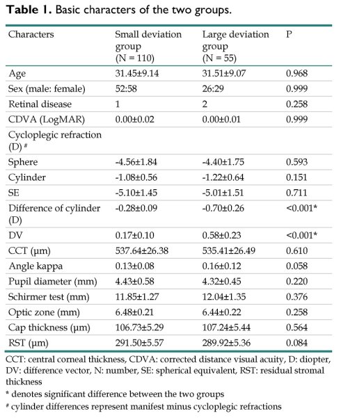
The preoperative characteristics of the two groups are summarized in Table 1. The mean age was 31.45±9.14 years in the small deviation group and 31.51±9.07 years in the large deviation group, with no significant between-group difference (P = 0.968). Sex distribution, and retinal disease ratio were also comparable between groups (both P > 0.05). Regarding ophthalmic parameters, CDVA, cycloplegic refraction, topographic measurements, and Schirmer test results did not differ significantly between the small deviation and large deviation groups (all P > 0.05). The difference of cylinder and DV was significantly greater in the large deviation group than the small deviation group (both P < 0.001). Surgical parameters were likewise comparable between groups (all P > 0.05) (Table 1).
Basic characters of the two groups.
| Characters | Small deviation group (N = 110) | Large deviation group (N = 55) | P |
|---|---|---|---|
| Age | 31.45±9.14 | 31.51±9.07 | 0.968 |
| Sex (male: female) | 52:58 | 26:29 | 0.999 |
| Retinal disease | 1 | 2 | 0.258 |
| CDVA (LogMAR) | 0.00±0.02 | 0.00±0.01 | 0.999 |
| Cycloplegic refraction (D) # | |||
| Sphere | -4.56±1.84 | -4.40±1.75 | 0.593 |
| Cylinder | -1.08±0.56 | -1.22±0.64 | 0.151 |
| SE | -5.10±1.45 | -5.01±1.51 | 0.711 |
| Difference of cylinder (D) | -0.28±0.09 | -0.70±0.26 | <0.001* |
| DV | 0.17±0.10 | 0.58±0.23 | <0.001* |
| CCT (μm) | 537.64±26.38 | 535.41±26.49 | 0.610 |
| Angle kappa | 0.13±0.08 | 0.16±0.12 | 0.058 |
| Pupil diameter (mm) | 4.43±0.58 | 4.32±0.45 | 0.220 |
| Schirmer test (mm) | 11.85±1.27 | 12.04±1.35 | 0.376 |
| Optic zone (mm) | 6.48±0.21 | 6.44±0.22 | 0.258 |
| Cap thickness (μm) | 106.73±5.29 | 107.24±5.44 | 0.564 |
| RST (μm) | 291.50±5.57 | 289.92±5.36 | 0.084 |
CCT: central corneal thickness, CDVA: corrected distance visual acuity, D: diopter, DV: difference vector, N: number, SE: spherical equivalent, RST: residual stromal thickness
* denotes significant difference between the two groups
# cylinder differences represent manifest minus cycloplegic refractions
At postoperative day 1, mean UDVA was similar between the small deviation group and the large deviation group (0.16±0.13 vs. 0.20±0.17, P = 0.096). However, at three months postoperatively, mean UDVA was better in the small deviation group (0.02±0.02 vs. 0.04±0.03, P < 0.001) (Table 2). Regarding refractive outcomes, SE was significantly greater in the large deviation group than the small deviation group at postoperative day 1 (-0.34±0.41 vs. -0.58±0.52, P = 0.002), and this difference persisted until the final follow-up visit (-0.12±0.06 vs. -0.25±0.15, P <0.001) (Table 2). At the final visit, 107 patients (97.27%) in the small deviation group and 50 patients (90.91%) in the large deviation group achieved 20/20 vision, with no significant difference between groups (P = 0.073) (Table 3). The proportions of patients with a loss of more than one line of CDVA was also similar between groups (P = 0.999) while proportions of patients with SE within ±0.50 D was significantly higher in the small deviation group than the large deviation group (P = 0.004) (Table 3). Vector analysis showed that DV (P <0.001), ME (P = 0.002) and CoI (P < 0.001) was significantly better in the small deviation group than in the large deviation group, whereas all other vector parameters were comparable between the two groups (all P > 0.05) (Table 4).
Postoperative visual acuity and spherical equivalent between the two groups.
| Outcome | Small deviation group (N = 110) | Large deviation group (N = 55) | P |
|---|---|---|---|
| UDVA (LogMAR) | |||
| 1 day | 0.16±0.13 | 0.20±0.17 | 0.096 |
| 1 week | 0.14±0.09 | 0.17±0.16 | 0.125 |
| 1 month | 0.06±0.03 | 0.09±0.05 | < 0.001* |
| 3 months | 0.02±0.02 | 0.04±0.03 | < 0.001* |
| SE | |||
| 1 day | -0.34±0.41 | -0.58±0.52 | 0.002* |
| 1 week | -0.13±0.17 | -0.30±0.27 | < 0.001* |
| 1 month | -0.12±0.10 | -0.26±0.19 | < 0.001* |
| 3 months | -0.12±0.06 | -0.25±0.15 | < 0.001* |
N: number, SE: spherical equivalent, UDVA: uncorrected distance visual acuity
* denotes significant difference between the two groups
The postoperative outcomes at the final visit between the two groups.
| Outcome (%) | Small deviation group (N = 110) | Large deviation group (N = 55) | P |
|---|---|---|---|
| UDVA (LogMAR) | 0.073 | ||
| < 20/20 | 3 (2.72%) | 5 (9.09%) | |
| ≥ 20/20 | 107 (97.28%) | 50 (90.91%) | |
| CDVA | 0.999 | ||
| No change or gain | 109 (99.09%) | 54 (98.18%) | |
| Loss ≥ 1 line | 1 (0.91%) | 1 (1.82%) | |
| SE | 0.004* | ||
| ≤ ±0.50D | 104 (94.55%) | 44 (80.00%) | |
| > ±0.50D | 6 (5.45%) | 11 (20.00%) |
CDVA: corrected distance visual acuity, N: number, SE: spherical equivalent, UDVA: uncorrected distance visual acuity
* denotes significant difference between the two groups
The vector analysis of astigmatism between the two groups.
| Parameter | Small deviation group (N = 110) | Large deviation group (N = 55) | P |
|---|---|---|---|
| TIA | 1.08±0.56 | 1.22±0.64 | 0.151 |
| SIA | 0.98±0.48 | 1.03±0.57 | 0.555 |
| DV | 0.24±0.17 | 0.41±0.29 | < 0.001* |
| ME | 0.10±0.14 | 0.19±0.23 | 0.002* |
| AE | 3.18±10.64 | 4.61±13.37 | 0.457 |
| CoI | 0.91±0.06 | 0.84±0.10 | < 0.001* |
AE: angle of error, CoI: correction index, DV: difference vector, ME: magnitude of error, N: number, SIA: surgically induced astigmatism, TIA: target-induced astigmatism
* denotes a significant difference between groups
For the association between preoperative factors and significant residual astigmatism, there was no significant correlation between all factors and significant residual astigmatism in the small deviation group (all P > 0.05) (Table 5). On the contrary, the high myopia (P =0.018) and high astigmatism (P < 0.001) were significantly associated with the significant residual astigmatism in the large deviation group (Table 5).
Factor for significant residual astigmatism between the two groups.
| Factor | P |
|---|---|
| Small deviation group | |
| Old age | 0.279 |
| High myopia | 0.095 |
| High astigmatism | 0.062 |
| Large angle kappa | 0.421 |
| Large deviation group | |
| Old age | 0.473 |
| High myopia | 0.018* |
| High astigmatism | <0.001* |
| Large angle kappa | 0.051 |
* denotes significant difference between the two groups
Discussion
In the present study, the postoperative SE, either by absolute value and percentage above threshold, were significantly worse in the large deviation group compared to the small deviation group. In addition, certain astigmatic parameters including the DV, ME and CoI were significantly worse in the large deviation group compared to the small deviation group. Besides, the high myopia and high astigmatism correlated to significant residual astigmatism in the large deviation group while no preoperative factor correlate to significant residual astigmatism in the small deviation group.
The mean postoperative SE and the percentage that SE above 0.50D were significantly higher in the large deviation group than the small deviation group. In the previous study assess the influence of astigmatism on the outcome of refractive surgery, the high astigmatism may associate with the higher risk of residual astigmatism after keratorefractive surgery [21]. Besides, the high corneal astigmatism was correlated to the higher incidence of re-treatment after keratorefractive surgery [22]. However, there was rare publication that evaluate the effect of the astigmatic deviation after cycloplegia procedure on the refractive outcome. To our knowledge, the findings of the present study may be a preliminary experience that demonstrate the association between large astigmatic deviation and the worse postoperative refractive outcome of second generation KLEx surgery. In addition, the baseline characteristics between the two groups, except for the astigmatic deviation degree after cycloplegia, were statistically similar thus the influence of other factors on the outcome assessment may be low. Furthermore, all the participants received identical preoperative and postoperative management and measurement because the Nobel Eye Institute arranged same postoperative protocol in all the branches and the operating error may not be prominent. Accordingly, the large astigmatic deviation may indeed be associated with the higher residual postoperative refractive error. Regarding the vector analysis, the DV, ME and CoI demonstrated significant worse values in the large deviation group than the small deviation group. On contrary, the SIA was similar between the two groups and the TIA differed by approximately 13% between the large and small deviation groups which is not a prominent difference. These results further demonstrate the moderate association between the large preoperative astigmatic deviation and the postoperative residual astigmatism in second generation KLEx surgery.
Concerning the correlation between the preoperative factors and the significant residual astigmatism in the small and large deviation groups, the high baseline myopia and high baseline astigmatism correlated to the significant residual astigmatism with statistical significance. The high myopia is a well-known predisposing factor for the suboptimal postoperative outcome of keratorefractive surgery [23]. In the previous study, the preoperative high myopia is correlated to the lower predictability of postoperative outcome [24]. In addition, the presence of high myopia may contribute to the postoperative residual myopia and astigmatism [22]. The high preoperative astigmatism is another established indicator for the guarded outcome of refractive surgery [25]. In the earlier literature, the high preoperative astigmatism is associated with poor refractive outcome of the first generation KLEx surgery [26]. Although the influence of preoperative high astigmatism was lower in the second generation KLEx compared to the first generation KLEx surgery [20], the influence could still exist and lead to lower refractive accuracy of second generation KLEx surgery. Consequently, it is reasonable that the high myopia and high astigmatism reduce the performance of second generation KLEx surgery in those with large astigmatic deviation after cycloplegia. Of note, the association between large angle kappa and significant residual astigmatism in large deviation population also reach marginal significance. On the other hand, there was no indicator for the significant residual astigmatism in the small deviation group. There was scant publication to report this phenomenon and difference. We think the possible reason is that the fair astigmatism correction ability of second generation KLEx surgery compensate the influence of risk factors in those without prominent astigmatic deviation. This results corresponded to the vector analysis results of the present study.
For the postoperative visual acuity and safety concern, the small deviation group represented a better postoperative UDVA than the large deviation group throughout the study period. The difference became significant after one month postoperatively. The residual astigmatism can influence the postoperative visual acuity in the previous publication [27], thus it is possible that the higher amount of residual astigmatism in the large deviation group reduced the postoperative UDVA compared to the small deviation population. Nevertheless, the difference of UDVA between the two groups may be statistically significant but not clinically significant. Except for the similar postoperative UDVA in the postoperative day one and postoperative week one, the UDVAs of the two groups one and three months postoperatively is near 20/20 and the 0.02 difference in LogMAR calculation could not influence the visual performance in the real world. Thus, the amplitude of residual astigmatism in the large deviation population may not affect the visual performance prominently. Moreover, the safety parameter could also be adequate for those with large astigmatic deviation since the number of participants who lost one or more lines of CDVA between group was statistical insignificant. The eyes that loss CDVA in both groups can still reach a CDVA of 0.05 LogMAR, which indicate that the astigmatic shift did not lead to prominent risk for the second generation KLEx surgery. Overall, the large astigmatic deviation may interfere the surgical outcome of second generation KLEx surgery but the degree may not be pronounced.
About the postoperative outcomes, the visual and refractive results of the two keratorefractive procedures assessed in the present study were generally comparable to those reported for other refractive surgical techniques. The postoperative UDVA of the large deviation group was 0.04, which is consistent with previously published outcomes for LASIK and FS-LASIK [28, 29]. Similarly, the 3-month postoperative UDVA observed in the small deviation group was 0.02, which was in line with that reported in a prior study evaluating the same procedure [30]. Regarding refractive accuracy, all eyes in the small deviation group achieved a postoperative SE within ±1.00 D, while 94.55% were within ±0.50 D. These findings are consistent with previously described refractive outcomes after both LASIK and KLEx procedures [28, 31]. Comparable results were also observed in the large deviation group, in which 100% and 80.00% of eyes achieved a postoperative SE within ±1.00 D and ±0.50 D, respectively. These results are in agreement with earlier reports on LASIK and first-generation KLEx [6, 9, 32]. Furthermore, the small deviation and large deviation groups yielded a safety index of 0.99 and 0.98, which was comparable to the safety index of 1.06 previously reported for LASIK [33]. Taken together, these findings indicate that the surgical performance of second-generation KLEx surgery at our institution was comparable to that reported in the existing literature regardless of the astigmatic deviation.
Several limitations should be acknowledged in the present study. First, the retrospective design may have introduced inherent selection bias and limited the completeness and consistency of data collection compared with a prospective study design. In particular, information on medication adherence, duration of visual display terminal use, and detailed contact lens-wearing history was not consistently available. Second, higher-order aberrations were not assessed either before or after surgery; therefore, changes in optical quality related to higher-order aberrations could not be evaluated, despite their relevance when comparing refractive surgical procedures. In addition, postoperative corneal topographic changes and axial length alterations were not measured. As these parameters may provide further insight into postoperative corneal morphology and refractive stability, their absence should be considered when interpreting the present findings. The follow-up period of our study is also relatively short in which corneal wound healing and epithelial remodeling after lenticule extraction can continue up to 6-12 months. A study for long-term stability assessments of KLEx surgery is needed. Finally, all the participants in the present study were Han Taiwanese, and the external validity of the present study could be reduced.
In conclusion, the large astigmatic deviation after cycloplegia may correlate with suboptimal refractive outcome of second generation KLEx surgery. Furthermore, the risk of significant residual astigmatism in this population could be higher if high refractive error existed. Consequently, repeated preoperative measurement of astigmatism in this population could be suggested. Further large-scale prospective study to evaluate the influence of cycloplegia astigmatic deviation on different refractive surgeries is mandatory.
Abbreviations
AE: angle of error; AXL: axial length; CCT: central corneal thickness; CDVA: corrected distance visual acuity; CoI: correction index; D: diopter; DV: difference vector; KLEx: keratorefractive lenticule extraction; ME: magnitude of error; N: number; RST: residual stromal thickness; SD: standard deviation; SE: spherical equivalent; SIA: surgically induced astigmatism; TIA: target-induced astigmatism; UDVA: uncorrected distance visual acuity.
Competing Interests
The authors have declared that no competing interest exists.
References
1. Ganesh S, Brar S, Arra RR. Refractive lenticule extraction small incision lenticule extraction: A new refractive surgery paradigm. Indian J Ophthalmol. 2018;66:10-9
2. Ang M, Gatinel D, Reinstein DZ, Mertens E, Alió Del Barrio JL, Alió JL. Refractive surgery beyond 2020. Eye (Lond). 2021;35:362-82
3. Padmanabhan P, Mrochen M, Basuthkar S, Viswanathan D, Joseph R. Wavefront-guided versus wavefront-optimized laser in situ keratomileusis: contralateral comparative study. J Cataract Refract Surg. 2008;34:389-97
4. Abdel-Radi M, Shehata M, Mostafa MM, Aly MOM. Transepithelial photorefractive keratectomy: a prospective randomized comparative study between the two-step and the single-step techniques. Eye (Lond). 2023;37:1545-52
5. Chang JY, Lin PY, Hsu CC, Liu CJ. Comparison of clinical outcomes of LASIK, Trans-PRK, and SMILE for correction of myopia. J Chin Med Assoc. 2022;85:145-51
6. Reinstein DZ, Carp GI, Archer TJ, Vida RS, Yammouni R. Large Population Outcomes of Small Incision Lenticule Extraction in Young Myopic Patients. J Refract Surg. 2022;38:488-96
7. Reinstein DZ, Archer TJ, Vida RS, Carp GI, Reinstein JFR, McAlinden C. Objective and Subjective Quality of Vision After SMILE for High Myopia and Astigmatism. J Refract Surg. 2022;38:404-13
8. Jabbarvand M, Khodaparast M, Moravvej Z, Shahraki K, Ahmadi H, Shahraki K. et al. Vector analysis of moderate to high myopic astigmatism after small-incision lenticule extraction (SMILE): 12-month follow-up. Eur J Ophthalmol. 2022;32:3312-20
9. Zhong Y, Li M, Han T, Fu D, Zhou X. Four-year outcomes of small incision lenticule extraction (SMILE) to correct high myopic astigmatism. Br J Ophthalmol. 2021;105:27-31
10. Zhang Y, Shen Q, Jia Y, Zhou D, Zhou J. Clinical Outcomes of SMILE and FS-LASIK Used to Treat Myopia: A Meta-analysis. J Refract Surg. 2016;32:256-65
11. Song J, Cao H, Chen X, Zhao X, Zhang J, Wu G. et al. Small Incision Lenticule Extraction (SMILE) Versus Laser Assisted Stromal In situ Keratomileusis (LASIK) for Astigmatism Corrections: A Systematic Review and Meta-analysis. Am J Ophthalmol. 2023;247:181-99
12. Hashemi H, Asgari S, Khabazkhoob M, Heidari Z. Vector analysis of astigmatism correction after PRK, FS-LASIK, and SMILE for myopic astigmatism. Int Ophthalmol. 2023;43:3999-4009
13. Lee JK, Chuck RS, Park CY. Femtosecond laser refractive surgery: small-incision lenticule extraction vs. femtosecond laser-assisted LASIK. Curr Opin Ophthalmol. 2015;26:260-4
14. Alió Del Barrio JL, Vargas V, Al-Shymali O, Alió JL. Small incision lenticule extraction (SMILE) in the correction of myopic astigmatism: outcomes and limitations - an update. Eye Vis (Lond). 2017;4:26
15. Zhao X, Zhang L, Ma J, Li M, Zhang J, Zhao X. et al. Comparison of Wavefront-Guided Femtosecond LASIK and Optimized SMILE for Correction of Moderate-to-High Astigmatism. J Refract Surg. 2021;37:166-73
16. Tian H, Gao W, Xu C, Wang Y. Clinical outcomes and higher order aberrations of wavefront-guided LASIK versus SMILE for correction of myopia: A systemic review and meta-analysis. Acta Ophthalmol. 2023;101:606-18
17. Saad A, Klabe K, Kirca M, Kretz FAT, Auffarth G, Breyer DRH. Refractive outcomes of small lenticule extraction (SMILE) Pro® with a 2 MHz femtosecond laser. Int Ophthalmol. 2024;44:52
18. Reinstein DZ, Archer TJ, Potter JG, Gupta R, Wiltfang R. Refractive and Visual Outcomes of SMILE for Compound Myopic Astigmatism with the VISUMAX 800. J Refract Surg. 2023;39:294-301
19. Brar S, Ganesh S, Bhargav S. Comparison of Intraoperative Time Taken for Docking, Lenticule Dissection, and Overall Workflow for SMILE Performed with the VisuMax 800 Versus the VisuMax 500 Femtosecond Laser. J Refract Surg. 2023;39:648
20. Lee CY, Lian LB, Chen HC, Huang CT, Huang JY, Yang SF. et al. The outcomes of first-generation (visumax 500) and second-generation (Visumax 800) keratorefractive lenticule extraction surgeries for astigmatism. Sci Rep. 2024;14:22224
21. Lee CY, Shen JH, Chao CC, Lian IB, Huang JY, Yang SF. et al. Topographic and surgical risk factors for high postoperative residual astigmatism after small incision lenticule extraction in patients with different degrees of myopia: a retrospective cohort study. BMC Ophthalmol. 2024;24:45
22. Muqbel Z, Förster A, Alkarkoukly S, Arba-Mosquera S, Dick HB, Taneri S. Risk Factors for Re-treatment After Keratorefractive Lenticule Extraction (KLEx) for Myopia and Myopic Astigmatism. J Refract Surg. 2024;40:e362-e70
23. Sommer A, Safir M, Santhiago MR, Nasser W, Ben Ephraim Noyman D, Sela T. et al. A Comparison of LASIK Outcomes for High Versus Low Myopia: Large Data Analysis. J Refract Surg. 2025;41:e797-e804
24. Lee C-Y, Yang S-F, Chen H-C, Lian I-B, Huang C-T, Huang J-Y. et al. The Risk Factors for Myopia Undercorrection in Second-Generation (Visumax 800) Keratorefractive Lenticule Extraction Surgery: A Retrospective Case-Control Study. Diagnostics. 2024;14:1752
25. Sorkin N, Kaiserman I, Kayal M, Levinger E, Mashour RS, Sela T. et al. Factors Predicting the Need for Re-treatment After Laser Refractive Surgery in Patients with Mixed Astigmatism. J Refract Surg. 2024;40:e73-e8
26. Lee CY, Jeng YT, Chao CC, Lian IB, Huang JY, Yang SF. et al. Refraction and topographic risk factors for early myopic regression after small-incision lenticule extraction surgery. Sci Rep. 2024;14:8732
27. Wu HK. Astigmatism and LASIK. Curr Opin Ophthalmol. 2002;13:250-5
28. Piñero DP, Teus MA. Clinical outcomes of small-incision lenticule extraction and femtosecond laser-assisted wavefront-guided laser in situ keratomileusis. J Cataract Refract Surg. 2016;42:1078-93
29. Li L, Xiong L, Wang Z. Comparison of clinical outcomes of a corneal wavefront- and topography-guided platforms for laser in situ keratomileusis on virgin eyes: an expanded cohort study. Int Ophthalmol. 2024;44:331
30. Yoo TK, Kim D, Kim JS, Kim HS, Ryu IH, Lee IS. et al. Comparison of early visual outcomes after SMILE using VISUMAX 800 and VISUMAX 500 for myopia: a retrospective matched case-control study. Sci Rep. 2024;14:11989
31. Reinstein DZ, Archer TJ, Vida RS, Carp GI, Reinstein JFR, McChesney T. et al. Small Incision Lenticule Extraction (SMILE) for the Correction of High Myopia with Astigmatism. J Refract Surg. 2022;38:262-71
32. Zou H, Wei X, Li L, Wei D, Mao H, Huang Y. et al. Comparison of objective visual quality between SMILE and FS-LASIK in moderate-to-high myopia. Front Med (Lausanne). 2024;11:1408516
33. Alió Del Barrio JL, Canto-Cerdán M, Bo M, Subirana N, Alió JL. Laser-assisted in situ keratomileusis long term outcomes in late adolescence. Eur J Ophthalmol. 2021;31:2307-12
Author contact
Corresponding authors: Chao-Kai Chang, MD, PhD, Address: Nobel Eye Institute, No. 13-5, Gongyuan Rd., Zhongzheng Dist., Taipei 100008, Taiwan, Tel: +886-2-2370-5666, Fax: +886-2-2375-4509, E-mail: chaokaihinet.net; Po-Hsien Huang, MD, Address: Kaohsiung Nobel Eye Institute, No. 106, Ziyou 2nd Rd., Zuoying District, Kaohsiung City, 813, Taiwan, Tel: +886-7-558-5678, Fax: +886-7-558-5658, E-mail: peterdigitalcom.