Impact Factor ISSN: 1449-1907




Int J Med Sci 2026; 23(12):3621-3630. doi:10.7150/ijms.137847 This issue Cite
Research Paper
Transcriptomic Alterations in Peri-Implantitis Tissues from Patients with Diabetes, Hypertension, and Osteoporosis: An Exploratory Candidate-Gene Study
Zhibin Liu1,†, Su-young Choi2,†, Ke Huang1, Chae Yeon Kim1, SunHo Ahn2, Hae-Won Park2, Eungyung Kim3, Hyunjin Kim1, Youngkyun Lee4, Myoung Ok Kim1,  , Yong-Gun Kim2,5,
1. Department of Animal Science and Biotechnology, Research Institute for Innovative Animal Science, Kyungpook National University, Sangju-si 37224, Gyeong Sang Buk-do, Republic of Korea.
2. Department of Periodontology, Kyungpook National University School of Dentistry, Daegu, Korea.
3. Department of Endodontics, School of Dentistry, University of Alabama at Birmingham, Birmingham, Alabama 35294.
4. Department of Biochemistry, School of Dentistry, Kyungpook National University, Daegu, Korea.
5. Center of Excellence in Precision Medicine and Digital Health, Department of Physiology, Faculty of Dentistry, Chulalongkorn University, Bangkok 10330, Thailand.
†Zhibin Liu and Su-young Choi contributed equally to this work.
Received 2026-5-15; Accepted 2026-10-10; Published 2026-10-16
Abstract
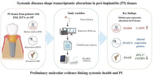
Background: Peri-implantitis (PI) is a significant cause of dental implant failure and is characterized by inflammatory destruction of peri-implant tissues with progressive bone loss. Increasing evidence indicates that systemic conditions such as diabetes mellitus (DM), hypertension (HTN), and osteoporosis (OP) may exacerbate peri-implant inflammation by affecting immune regulation and bone metabolism. However, the molecular mechanisms linking these diseases to PI remain insufficiently understood.
Methods: Peri-implant gingival tissues were collected from 50 patients treated at Kyungpook National University Dental Hospital and classified into healthy implant (HI) and PI groups. Exploratory RNA sequencing was performed to identify genes showing prominent expression changes between PI and HI tissues, followed by literature-informed candidate-gene selection and quantitative real-time PCR (RT-qPCR) validation.
Results: Transcriptomic analysis revealed distinct gene-expression patterns in PI tissues from patients with HTN, DM, or OP. Literature-informed prioritization identified nine HTN-related, fifteen DM-related, and four OP-related candidate genes. RT-qPCR validation confirmed several expression changes identified in the exploratory RNA sequencing screening. Strikingly, additional comparison with systemically healthy PI patients further revealed significant differences in CASP4 in PI-DM tissues and RPL17 and FGFBP1 in PI-OP tissues.
Conclusions: These findings identify distinct molecular alterations associated with PI in patients with HTN, DM, or OP and provide candidate genes for further investigation of the interplay between systemic health and peri-implant disease.
Keywords: peri-implantitis, diabetes mellitus, hypertension, osteoporosis, rna sequencing, gene expression
1. Introduction
Dental implants are widely regarded as an effective and predictable treatment option for replacing missing teeth and restoring oral function [1, 2]. However, peri-implantitis (PI) remains a significant biological complication that can lead to implant failure. PI is characterized by inflammatory destruction of peri-implant tissues accompanied by progressive loss of supporting bone, leading to functional and aesthetic compromise. Its pathogenesis is multifactorial, involving microbial biofilm accumulation, host immune responses, and genetic susceptibility [3, 4]. Although conventional therapeutic strategies such as mechanical debridement and antimicrobial interventions can alleviate clinical symptoms, recurrence and chronic inflammation are common [5]. These findings suggest that factors beyond local infection, including systemic conditions and transcriptomic alterations, significantly influence PI initiation and progression [6, 7].
Systemic conditions such as diabetes mellitus (DM), hypertension (HTN), and osteoporosis (OP) are increasingly recognized as potential modifiers for peri-implant health [8, 9]. DM is associated with impaired wound healing, enhanced inflammatory responses, and altered bone metabolism, which can compromise osseointegration and increase PI susceptibility [8]. HTN has been linked to microvascular dysfunction, potentially impairing tissue perfusion and delaying peri-implant healing [10]. OP disrupts bone remodeling and reduces bone density, predisposing peri-implant sites to enhanced bone resorption [9]. Although each of these conditions has been associated with adverse implant outcomes, the molecular mechanisms connecting systemic disease to PI remain insufficiently defined. Clarifying these mechanisms may explain interindividual differences in disease susceptibility.
Advances in transcriptomic technologies, particularly RNA sequencing (RNA-seq), enable transcriptome-wide profiling of gene-expression changes and have been widely applied to characterize molecular alterations associated with pathological processes, including inflammatory diseases of oral tissues [11-13]. Analysis of peri-implant tissues may facilitate the identification of molecular alterations and candidate biomarkers associated with PI [14, 15]. Integrating transcriptomic data with clinical characteristics further enhances understanding of how systemic disorders modulate the local peri-implant tissue environment and immune response [16]. Previous transcriptomic studies have explored potential molecular links between PI and systemic diseases. For example, Yu et al. identified shared genes and signaling pathways associated with PI and type 2 diabetes mellitus through integrative bioinformatics analysis of publicly available transcriptomic datasets [17]. However, direct transcriptomic profiling of peri-implant tissues from patients with different systemic comorbidities, particularly hypertension and osteoporosis, remains limited.
Therefore, this study aimed to explore gene-expression alterations in PI tissues from patients with DM, HTN, or OP and to identify candidate genes potentially associated with the interplay between systemic conditions and PI. Exploratory transcriptomic screening was followed by literature-informed candidate-gene prioritization and RT-qPCR validation using independent biological samples.
2. Materials and Methods
2.1. Ethical statement
The study protocol was reviewed and approved by the Ethics Committee of Kyungpook National University Dental Hospital (Approval Nos.: KNUDH-2022-07-02-00, KNUDH-2024-04-05-00, and KNUDH-2025-05-05-00). All participants provided written informed consent prior to enrollment.
2.2. Patients and clinical evaluation
A total of 50 patients were enrolled, comprising 10 individuals with healthy implants (HI) and 40 patients diagnosed with PI. The PI cohort was divided into four subgroups—PI, PI with DM (PI-DM), PI with HTN (PI-HTN), and PI with OP (PI-OP)—each comprising 10 patients, regardless of sex. The median age of the study population was 66.0 years (interquartile range [IQR], 58.5-70.0 years). All participants underwent implant placement or PI treatment at Kyungpook National University Dental Hospital.
Comprehensive medical histories and clinical background data were obtained from all participants following informed consent to ensure accurate classification. Based on systemic disease status and peri-implant condition, participants were categorized into five groups: HI, PI, PI-DM, PI-HTN, and PI-OP. Patients in the HI group had healthy implants and no documented systemic diseases based on their medical histories and clinical records. DM, HTN, and OP were identified based on documented diagnoses in the patients' medical records and ongoing disease-specific treatment. Detailed classification criteria, sample sizes, age distributions, and baseline characteristics are summarized in Table S1.
The transcriptomic screening and quantitative real-time PCR (RT-qPCR) validation were performed at different analytical levels. For the exploratory RNA-seq analysis, RNA samples from three individuals within each group were pooled in equal amounts before library preparation, resulting in one pooled RNA-seq library per group. For subsequent RT-qPCR validation, gene expression was evaluated using independent biological samples from 10 patients in each group.
2.3. Library preparation and sequencing
Total RNA was extracted from peri-implant gingival tissues using a commercial RNA isolation kit (catalog number TS200-001; Tri-Solution, Bio Science Technology, Daegu, Republic of Korea). RNA purity was assessed spectrophotometrically, with 260/280 ratios of approximately 2.0 considered acceptable. For the pooled RNA samples used for RNA-seq, RNA integrity was additionally assessed using TapeStation analysis before library preparation, and RNA integrity number equivalent (RINe) values were recorded as part of the RNA quality-control procedure. The RINe values of the pooled RNA-seq samples ranged from 3.9 to 8.9.
For exploratory transcriptomic screening, RNA obtained from three individual participants within each group was pooled in equal amounts before library preparation, generating one pooled RNA-seq sample for each of the HI, PI-DM, PI-HTN, and PI-OP groups. The pooled RNA samples were processed by eBiogen Inc. (Seoul, Republic of Korea) for library preparation and sequencing.
RNA-seq data processing and exploratory expression profiling were performed using ExDEGA software (eBiogen Inc.). Expression data were normalized using trimmed mean of M-values (TMM) and counts per million (CPM) normalization. Genes showing prominent expression changes between each PI subgroup and the HI group were ranked according to fold-change magnitude and used for exploratory candidate-gene screening and visualization.
Because the three individual RNA samples within each group were pooled before library preparation, only one RNA-seq library was available per group. Therefore, the RNA-seq dataset did not contain independent biological replicates at the sequencing-library level, precluding reliable estimation of within-group biological dispersion and formal replicate-based differential-expression testing with multiple-testing correction. Accordingly, the RNA-seq results were interpreted as exploratory expression changes rather than statistically significant differentially expressed genes. Candidate genes identified through this exploratory transcriptomic screening were subsequently evaluated by RT-qPCR using independent biological samples.
2.4. RT-qPCR analysis
For the individual RNA samples used for RT-qPCR validation, RNA concentration and spectrophotometric purity were assessed prior to reverse transcription. Total RNA was reverse-transcribed into complementary DNA (cDNA) using the PrimeScript™ First Strand cDNA Synthesis Kit (Cat. No. 6110; Takara Bio Inc., Kyoto, Japan) with oligo-dT primers. Quantitative PCR (qPCR) was performed in 20-μL reaction volumes containing 8 μL of diluted cDNA, 10 μL of Power SYBR® Green PCR Master Mix (Cat. No. 4367659; Applied Biosystems, Thermo Fisher Scientific Inc., Waltham, Massachusetts, United States), and 1 μL each of gene-specific forward and reverse primers. β-actin (ACTB) was used as the endogenous reference gene for normalization. Primer sequences for ACTB and all target genes are provided in Table S2. All RT-qPCR assays were performed under identical amplification conditions. Post-amplification melting-curve analysis was conducted to assess product specificity, and only reactions showing specific amplification were included in the analysis. For each sample, ΔCt was calculated as Ct(target gene) - Ct(β-actin), and relative expression was calculated as 2^-ΔCt. The resulting values were then normalized to the mean 2^-ΔCt value of the HI control group, which was set to 1, and expression levels in the PI groups were expressed relative to the HI group.
2.5. Statistical analysis
Statistical analyses of clinical variables and RT-qPCR validation data were performed using GraphPad Prism 9 (GraphPad Software, San Diego, CA, USA). For RT-qPCR analysis, each patient was treated as an independent biological replicate (n = 10 per group), and the mean of technical replicates was used as the patient-level value. Normality was assessed using the Shapiro-Wilk test, with Q-Q plots used for graphical assessment of data distribution (Figure S1). Normally distributed data with equal variances were analyzed using ordinary one-way ANOVA followed by Tukey's multiple-comparisons test, whereas data with unequal variances were analyzed using Welch's ANOVA followed by Dunnett's T3 test. Non-normally distributed data were analyzed using the Kruskal-Wallis test followed by Dunn's multiple-comparisons test. All tests were two-sided, and adjusted p values < 0.05 were considered statistically significant. The exploratory RNA-seq screening was not subjected to inferential statistical testing because only one pooled sequencing library was available per group.
3. Results
3.1 Exploratory transcriptomic profiling of PI under systemic disease conditions
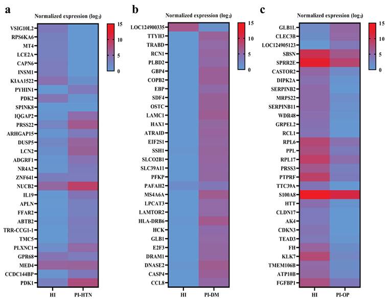
To explore gene expression alterations associated with PI in patients with different systemic conditions, RNA-seq was used as an exploratory transcriptomic screening approach. For each group, RNA from three individual tissue samples was pooled before sequencing, resulting in one pooled RNA-seq library per group. Expression profiles of the PI-HTN, PI-DM, and PI-OP groups were separately compared with that of the HI group. Heatmaps were generated using ExDEGA to visualize the top 30 genes showing the largest expression changes based on fold change magnitude in each comparison (Figure 1). The normalized expression values underlying the three heatmaps are provided in Table S3. The three PI subgroups showed different patterns of gene expression changes relative to the HI group, providing candidate genes for subsequent investigation.
Exploratory RNA-seq profiling of gene expression changes in PI associated with systemic conditions. Heatmaps show the top 30 genes exhibiting the largest expression changes based on fold-change magnitude in (a) PI-HTN versus HI, (b) PI-DM versus HI, and (c) PI-OP versus HI. Raw read counts were normalized using the trimmed mean of M-values (TMM) method, converted to counts per million (CPM), and log2-transformed for visualization. Blue and red indicate lower and higher normalized expression levels, respectively. All three heatmaps were visualized using the same color scale (0-15) based on log₂-transformed normalized expression values to permit direct visual comparison across disease groups. Hierarchical clustering was not applied; genes were displayed according to the magnitude of their expression changes in each comparison. For each group, RNA from three individual tissue samples was pooled before library preparation, resulting in one pooled RNA-seq library per group. Therefore, the heatmaps represent pooled group-level expression profiles and were used for exploratory visualization and candidate-gene screening rather than evaluation of inter-patient variability. Abbreviations: HI, healthy implant; HTN, hypertension; DM, diabetes mellitus; OP, osteoporosis.
To prioritize candidate genes for subsequent validation, a literature-informed selection strategy was applied to genes showing prominent expression changes in the exploratory RNA-seq screening. Genes were considered candidates when they had previously reported relevance to the corresponding systemic condition or related biological processes (HTN, DM, or OP). Nine candidate genes with previously reported relevance to hypertension, blood-pressure regulation, vascular biology, or related metabolic processes were identified (PDK2, DUSP5, LCN2, NR4A2, NUCB2, FFAR2, ABTB2, PLXNC1, and PDK1; Table 1). For DM, fifteen candidate genes with reported associations were identified (RCN1, SDF4, OSTC, LAMC1, EIF2S1, SLCO2B1, PFKP, MS4A6A, LAMTOR2, HCK, GLB1, E2F3, DRAM1, CASP4, and CCL8; Table 2). Four candidate genes with reported associations with OP were identified (CLEC3B, RPL17, TEAD3, and FGFBP1; Table 3).
Candidate genes prioritized from the PI-HTN RNA-seq screening based on literature-supported relevance to hypertension or related biological processes.
| Gene | Associated risk or condition | References |
|---|---|---|
| PDK2 | Vascular smooth muscle metabolic remodeling and proliferative vascular disease | [41] |
| DUSP5 | Hypertension-associated renal vascular injury | [42] |
| LCN2 | Essential hypertension and blood-pressure regulation | [28, 29] |
| NR4A2 | Atherosclerosis | [43] |
| NUCB2 | HTN | [44, 45] |
| FFAR2 | HTN | [46] |
| ABTB2 | Bronchial asthma and arterial HTN | [47] |
| PLXNC1 | HTN | [48] |
| PDK1 | Vascular endothelial metabolic reprogramming and inflammation | [30] |
Abbreviations: HTN, hypertension
Candidate genes prioritized from the PI-DM RNA-seq screening based on literature-supported relevance to diabetes or related biological processes.
| Gene | Associated risk or condition | References |
|---|---|---|
| RCN1 | Endoplasmic reticulum stress and diabetic foot | [49] |
| SDF4 | Diabetic foot | [50] |
| OSTC | Type 1 diabetes-associated transcriptional alteration | [51] |
| LAMC1 | Diabetic kidney disease | [34, 35] |
| EIF2S1 | Type 2 DM | [17] |
| SLCO2B1 | Diabetic kidney disease-associated expression alteration | [52] |
| PFKP | Diabetic kidney disease | [53] |
| MS4A6A | Nonalcoholic fatty liver disease, periodontitis, and type 2 DM | [33] |
| LAMTOR2 | Diabetic kidney disease | [54] |
| HCK | Diabetic nephropathy-associated immune/inflammatory processes | [55, 56] |
| GLB1 | Type 2 DM | [57] |
| E2F3 | Diabetic kidney disease | [58] |
| DRAM1 | Glucose-responsive autophagy/apoptosis regulation | [59] |
| CASP4 | Diabetic kidney disease | [60] |
| CCL8 | Diabetes-related inflammation | [61, 62] |
Abbreviations: DM, diabetes mellitus
Candidate genes prioritized from the PI-OP RNA-seq screening based on literature-supported relevance to osteoporosis or related biological processes.
| Gene | Associated risk or condition | References |
|---|---|---|
| CLEC3B | Inhibition of osteoclast differentiation and bone resorption | [38] |
| RPL17 | OP and COVID-19 | [63] |
| TEAD3 | Promotion of osteoclast differentiation | [40] |
| FGFBP1 | Altered bone mineral density | [39] |
Abbreviations: OP, osteoporosis; COVID-19, coronavirus disease 2019
3.2. Validation of systemic disease-associated genes by RT-qPCR
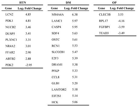
Based on RNA-seq results, linear fold-change values were converted to log₂ fold-change (log₂FC) values and used to identify genes with the most pronounced expression differences (Table 4). From these literature-supported candidate-gene lists, genes showing the most prominent expression changes were selected for RT-qPCR validation in each systemic disease subgroup. Specifically, three HTN-associated genes (LCN2, PDK1, and NUCB2), three DM-associated genes (MS4A6A, LAMC1, and CASP4), and four OP-associated genes (CLEC3B, RPL17, FGFBP1, and TEAD3) were examined using RT-qPCR. Moreover, to further distinguish gene-expression changes associated with systemic conditions from those related to peri-implant inflammation alone, we additionally included samples from systemically healthy patients with PI for comparison.
Log₂ fold changes of candidate genes prioritized in the PI-HTN, PI-DM, and PI-OP RNA-seq comparisons.
Abbreviations: HTN, hypertension; PI, peri-implantitis; HI, healthy implants; DM, diabetes mellitus; OP, osteoporosis; RT-qPCR, reverse transcription-polymerase chain reaction.
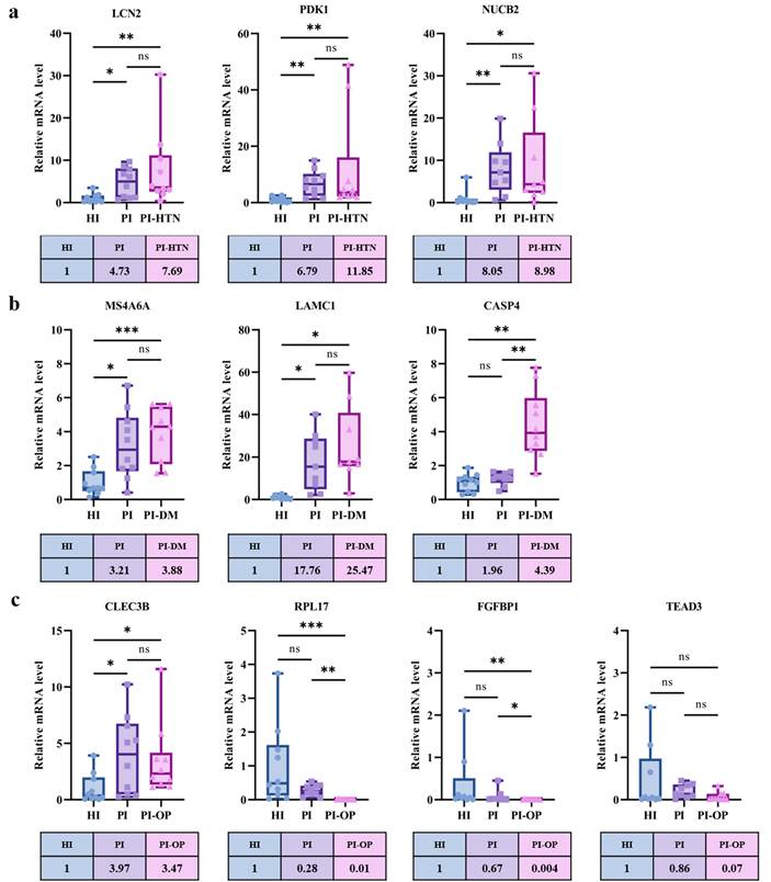
As shown in Figure 2, RT-qPCR analysis generally showed expression patterns directionally consistent with the exploratory RNA-seq screening. In the PI-HTN subgroup, expression of LCN2, PDK1, and NUCB2 was significantly increased relative to HI controls. Although the mean NUCB2 expression was numerically higher in the PI-HTN group than in the systemically healthy PI group (8.98 versus 8.05), the difference was not statistically significant. Similarly, MS4A6A, LAMC1, and CASP4 were substantially upregulated in PI-DM tissues. In the PI-OP subgroup, CLEC3B expression was significantly elevated, whereas RPL17 and FGFBP1 were significantly downregulated compared with HI tissues. However, CLEC3B expression was not higher in the PI-OP group than in the systemically healthy PI group (3.47 versus 3.97), and the difference was not statistically significant. TEAD3 showed a downward trend, but the difference was not statistically significant. Importantly, comparison with the systemically healthy PI group showed that CASP4 expression was significantly higher in PI-DM tissues than in PI tissues. In the OP-associated analysis, RPL17 and FGFBP1 also showed significant differences between PI-OP and systemically healthy PI tissues. These validation results confirm the RNA-seq-derived expression patterns and suggest that dysregulation of systemic disease-associated genes may contribute to increased susceptibility to PI in patients with DM or OP.
Validation of systemic disease-associated candidate genes in PI using quantitative RT-qPCR. (a) Expression of selected HTN-associated candidate genes in HI, systemically healthy PI, and PI-HTN tissues. (b) Expression of selected DM-associated candidate genes in HI, systemically healthy PI, and PI-DM tissues. (c) Expression of selected OP-associated candidate genes in HI, systemically healthy PI, and PI-OP tissues. Candidate genes were prioritized from the exploratory RNA-seq screening based on prominent expression changes and previously reported associations with the corresponding systemic conditions. Data are presented as box-and-whisker plots, with the center line indicating the median, the box representing the interquartile range (25th-75th percentiles), and the whiskers extending from the minimum to the maximum value. Each data point represents an independent biological sample (n = 10 per group). Relative mRNA levels were normalized to ACTB and are presented relative to the mean value of the HI control group, which was set to 1. Data were analysed using the statistical tests described in the Methods. Adjusted P values are indicated as *p < 0.05, ** p < 0.01, and *** p < 0.001. Abbreviations: HTN, hypertension; PI, peri-implantitis; HI, healthy implants; DM, diabetes mellitus; OP, osteoporosis; RT-qPCR, reverse transcription-polymerase chain reaction.
4. Discussion
PI remains a challenging biological complication in implant dentistry, jeopardizing long-term implant stability and patient quality of life. It is characterized by progressive inflammatory destruction of peri-implant soft tissues and supporting bone, frequently leading to discomfort, functional impairment, and eventual implant loss [4, 18]. Although bacterial biofilm is widely recognized as the primary etiological factor, increasing evidence suggests that host-related factors—particularly systemic diseases—considerably influence individual susceptibility and disease severity [19, 20]. Therefore, elucidating the biological mechanisms linking systemic health and peri-implant inflammation is essential for improving risk stratification, early diagnosis, and targeted intervention strategies [21].
Systemic conditions such as HTN, DM, and OP profoundly affect immune regulation, microvascular function, and bone metabolism, all of which are central to peri-implant tissue homeostasis [6, 21]. HTN is associated with vascular dysfunction and heightened oxidative stress, potentially impairing peri-implant soft tissue healing [22, 23]. DM promotes hyperinflammatory responses, alters neutrophil function, impedes collagen maturation, and disrupts osteoblast-osteoclast balance, thereby increasing susceptibility to peri-implant bone loss [8, 24, 25]. OP, characterized by reduced bone density and accelerated bone remodeling, compromises the structural support required for implant stability [26, 27]. Despite these established clinical associations, the molecular mechanisms linking systemic disorders to PI susceptibility remain incompletely defined. The present transcriptomic analysis offers mechanistic insight by identifying systemic disease-associated gene signatures within PI tissues.
In the PI-HTN subgroup (Figure 2), LCN2, PDK1, and NUCB2 were significantly upregulated relative to HI tissues. Although gene expression was numerically higher in the PI-HTN group than in the systemically healthy PI group, the difference was not statistically significant. Therefore, the increase in gene expression cannot be specifically attributed to HTN and may instead reflect PI-associated alterations. LCN2 (lipocalin-2) has been associated with vascular endothelial function and blood pressure regulation in patients with hypertension [28, 29]. Its upregulation in PI-HTN tissues may reflect inflammatory or vascular alterations, although its specific role in PI remains to be established. PDK1, a key metabolic regulator, has been implicated in vascular endothelial metabolic reprogramming and inflammation [30], suggesting that altered local metabolism may contribute to the inflammatory environment in PI-HTN tissues. NUCB2 (nesfatin-1 precursor) participates in blood pressure regulation and inflammatory signaling [31, 32]. Collectively, these findings suggest that altered inflammatory, metabolic, and vascular processes may contribute to the molecular characteristics observed in PI tissues.
In the DM subgroup, MS4A6A, LAMC1, and CASP4 displayed significant upregulation in PI tissues. MS4A6A has been implicated in chronic inflammatory states and macrophage dysregulation, both of which are prominent features of diabetic tissues [33]. LAMC1, a component of the extracellular matrix, has been implicated in diabetic retinopathy and hyperglycemia-associated endothelial alterations [34, 35]. Its upregulation in PI-DM tissues may be associated with changes in extracellular matrix organization and local tissue responses under diabetic conditions. CASP4, a central regulator of noncanonical inflammasome activation, mediates pyroptosis and chronic inflammatory signaling in metabolic disorders [36, 37]. Notably, the additional comparison with systemically healthy PI patients showed that CASP4 expression was significantly higher in the PI-DM group. Because both groups had PI but differed in DM status, this finding suggests that the increase in CASP4 expression may not be attributable to PI alone and may be associated with the presence of DM. These expression patterns suggest that altered inflammatory responses and tissue-remodeling processes may contribute to the molecular characteristics of PI in patients with DM.
Distinct transcriptomic alterations were also observed in the OP subgroup. CLEC3B, which encodes tetranectin (TNA), has been reported to inhibit osteoclast differentiation and bone resorption [38]. However, CLEC3B expression was not higher in the PI-OP group than in the systemically healthy PI group, suggesting that its increased expression may reflect PI-associated alterations rather than an OP-specific effect. Conversely, RPL17 and FGFBP1 were downregulated in PI-OP tissues. FGFBP1 modulates fibroblast growth factor signaling with known implications in bone density maintenance, and its reduced expression may compromise local osteogenic potential [39]. TEAD3 has been reported to promote osteoclast differentiation through activation of NFATC1 [40]. However, the observed difference in TEAD3 expression was not statistically significant, and further studies with larger sample sizes are needed to confirm this expression pattern. Importantly, comparison with systemically healthy PI patients revealed significant differences in RPL17 and FGFBP1 expression in the PI-OP group. Because both groups had PI, these differences suggest that the expression patterns of RPL17 and FGFBP1 may be associated with the presence of OP rather than reflecting PI alone. These findings are consistent with a potential link between altered skeletal metabolism in OP and increased peri-implant bone vulnerability.
Finally, several limitations of this study should be acknowledged. Although several candidate genes showed directional expression differences, most did not reach statistical significance when PI patients with systemic diseases were compared with systemically healthy PI patients. The relatively small sample size of each systemic disease subgroup may have limited the generalizability of our findings and reduced the statistical power to detect subtle molecular differences. In addition, detailed clinical information regarding glycemic control, including HbA1c levels, blood pressure control, and antihypertensive drug classes, as well as bone mineral density T-scores and antiresorptive medication use, was not consistently available for all participants. Consequently, stratified analyses according to disease severity, systemic disease control, or medication use could not be performed. These clinical factors may influence tissue homeostasis, inflammatory responses, bone metabolism, and local gene expression and should therefore be considered potential confounders. Future studies incorporating standardized clinical measurements and detailed medication histories are needed to determine their independent contributions to the gene-expression patterns observed in peri-implant tissues. Although the inclusion of systemically healthy PI patients helped distinguish gene-expression changes potentially associated with systemic conditions from those related to PI alone, HI tissue samples from individuals with DM, HTN, or OP were not available. Therefore, the independent effects of systemic diseases and their potential interactions with PI could not be fully disentangled. Future studies involving larger, age-matched cohorts and multivariable analyses are needed to determine whether these candidate genes are independently associated with systemic disease-related susceptibility to PI.
5. Conclusion
This study identified distinct gene-expression profiles in PI tissues from patients with HTN, DM, and OP and highlighted candidate genes potentially linking systemic conditions to the local molecular environment of PI. RNA-seq screening followed by RT-qPCR validation demonstrated consistent alterations in selected candidate genes, while additional comparisons with systemically healthy PI patients further revealed significant differences in CASP4 in the PI-DM group and RPL17 and FGFBP1 in the PI-OP group. These findings provide preliminary evidence that selected molecular alterations, particularly CASP4 in PI-DM and RPL17 and FGFBP1 in PI-OP, may be associated with systemic comorbidities beyond changes related to PI alone. Overall, our findings provide new molecular insights into the interplay between systemic health and PI and identify candidate molecular markers for further investigation of disease susceptibility and individualized risk assessment.
Supplementary Material
Supplementary figure S1 and tables.

Acknowledgements
Funding statement
This work was supported by the National Research Foundation of Korea (NRF) grant funded by the Korea government (MSIT) (RS-2025-00559717, Y-G K). This research was also supported by the Regional Innovation System & Education (RISE) Glocal 30 program through the Daegu RISE Center, funded by the Ministry of Education (MOE) and the Daegu, Republic of Korea (2025-RISE-03-001).
Author contributions
Z.B.L.: Conceptualization; data curation; validation; writing - review and editing. S.Y.C.: Conceptualization; data curation; validation; writing - review and editing. K.H.: Writing - original draft. C.Y.K.: Data curation; software. S.H.A.: Writing - original draft. H.W.P.: Data curation; software. E.Y.K.: Formal analysis. H.J.K.: Visualization. Y.K.L.: Visualization; formal analysis; resources. M.O.K.: Conceptualization; resources; supervision; review and editing. Y.G.K.: Resources; project administration; funding acquisition; review.
Data availability statement
Due to ethical restrictions concerning human-derived sequencing data, the raw RNA sequencing data cannot be made publicly available. The TMM- and CPM-normalized expression data underlying Figure 1 are provided in Table S3.
ORCID
Zhibin Liu: https://orcid.org/0000-0002-0444-4819
Su-young Choi: https://orcid.org/0009-0003-4646-9058
Ke Huang: https://orcid.org/0000-0001-7421-662X
Chae Yeon Kim: https://orcid.org/0000-0001-7301-4444
SunHo Ahn: https://orcid.org/0009-0002-5453-1870
Hae-Won Park: https://orcid.org/0009-0008-1869-0130
Eungyung Kim: https://orcid.org/0000-0001-8434-0915
Hyunjin Kim: https://orcid.org/0000-0002-6553-3524
Youngkyun Lee: https://orcid.org/0000-0001-9049-6908
Myoung Ok Kim: https://orcid.org/0000-0001-6650-7734
Yong-Gun Kim: https://orcid.org/0000-0002-2793-7667
Competing Interests
The authors have declared that no competing interest exists.
References
1. Guillaume B. Dental implants: A review. Morphologie. 2016;100:189-98
2. Addy LD. An introduction to dental implants. Br Dent J. 2024;236:753-7
3. Darby I. Risk factors for periodontitis & peri-implantitis. Periodontol 2000. 2022;90:9-12
4. Schwarz F, Derks J, Monje A, Wang HL. Peri-implantitis. J Periodontol. 2018;89(Suppl 1):S267-s90
5. Chambrone L, Wang HL, Romanos GE. Antimicrobial photodynamic therapy for the treatment of periodontitis and peri-implantitis: An American Academy of Periodontology best evidence review. J Periodontol. 2018;89:783-803
6. Jepsen S, Caton JG, Albandar JM, Bissada NF, Bouchard P, Cortellini P. et al. Periodontal manifestations of systemic diseases and developmental and acquired conditions: Consensus report of workgroup 3 of the 2017 World Workshop on the Classification of Periodontal and Peri-Implant Diseases and Conditions. J Periodontol. 2018;89(Suppl 1):S237-s48
7. Săndulescu M, Sîrbu VD, Popovici IA. Bacterial species associated with peri-implant disease - a literature review. Germs. 2023;13:352-61
8. Wagner J, Spille JH, Wiltfang J, Naujokat H. Systematic review on diabetes mellitus and dental implants: an update. Int J Implant Dent. 2022;8:1
9. Insua A, Galindo-Moreno P, Miron RJ, Wang HL, Monje A. Emerging factors affecting peri-implant bone metabolism. Periodontol 2000. 2024;94:27-78
10. Hamadé L, El-Disoki S, Chrcanovic BR. Hypertension and Dental Implants: A Systematic Review and Meta-Analysis. J Clin Med. 2024;13:499
11. Conesa A, Madrigal P, Tarazona S, Gomez-Cabrero D, Cervera A, McPherson A. et al. A survey of best practices for RNA-seq data analysis. Genome Biol. 2016;17:13
12. Gurgel BCV, Vilela N, Soares KH, Juliasse LER, Aukhil I, Duarte PM. Comprehensive analysis of transcriptome and pathway interactions in periodontitis. J Appl Oral Sci. 2025;33:e20250396
13. Lundmark A, Gerasimcik N, Båge T, Jemt A, Mollbrink A, Salmén F. et al. Gene expression profiling of periodontitis-affected gingival tissue by spatial transcriptomics. Sci Rep. 2018;8:9370
14. Zhang H, Zhang X, Huang J, Fan X. Identification of key genes and pathways for peri-implantitis through the analysis of gene expression data. Exp Ther Med. 2017;13:1832-40
15. Li Q, Lu H, Zhang M, Ye Y, Chen Q, Sun P. Epigenetic factors associated with peri-implantitis: a review. J Zhejiang Univ Sci B. 2025;26:657-74
16. Dionigi C, Ericson AC, Shikhan A, Larsson L, Berglundh T. Spatial Transcriptomic Assessments of Gene Expression Profiles in Human Peri-Implant Lesions. J Clin Periodontol. 2025;53(1):137-51
17. Yu T, Acharya A, Mattheos N, Li S, Ziebolz D, Schmalz G. et al. Molecular mechanisms linking peri-implantitis and type 2 diabetes mellitus revealed by transcriptomic analysis. PeerJ. 2019;7:e7124
18. Roccuzzo A, Imber JC, Salvi GE, Roccuzzo M. Peri-implantitis as the consequence of errors in implant therapy. Periodontol 2000. 2023;92:350-61
19. Schwarz F, Derks J, Monje A, Wang HL. Peri-implantitis. J Clin Periodontol. 2018;45(Suppl 20):S246-s66
20. Jepsen S, Berglundh T, Genco R, Aass AM, Demirel K, Derks J. et al. Primary prevention of peri-implantitis: managing peri-implant mucositis. J Clin Periodontol. 2015;42(Suppl 16):S152-7
21. Chapple ILC, Mealey BL, Van Dyke TE, Bartold PM, Dommisch H, Eickholz P. et al. Periodontal health and gingival diseases and conditions on an intact and a reduced periodontium: Consensus report of workgroup 1 of the 2017 World Workshop on the Classification of Periodontal and Peri-Implant Diseases and Conditions. J Periodontol. 2018;89(Suppl 1):S74-s84
22. Fu D, Shu X, Zhou G, Ji M, Liao G, Zou L. Connection between oral health and chronic diseases. MedComm (2020). 2025;6:e70052
23. Virdis A, Duranti E, Taddei S. Oxidative Stress and Vascular Damage in Hypertension: Role of Angiotensin II. Int J Hypertens. 2011;2011:916310
24. Wu YY, Xiao E, Graves DT. Diabetes mellitus related bone metabolism and periodontal disease. Int J Oral Sci. 2015;7:63-72
25. Portes J, Bullón B, Quiles JL, Battino M, Bullón P. Diabetes Mellitus and Periodontitis Share Intracellular Disorders as the Main Meeting Point. Cells. 2021;10:2411
26. Lee SA, Yi YJ, Won S, Chang NH, Kim JH. Prognostic factors influencing implant survival and marginal bone loss in patients with osteoporosis or osteopenia medication. J Korean Assoc Oral Maxillofac Surg. 2025;51:17-25
27. Liu F, Wang X, He Y, Han R, Wang T, Guo Y. Jaw osteoporosis: Challenges to oral health and emerging perspectives of treatment. Biomedicine & Pharmacotherapy. 2024;177:116995
28. Park CG, Choi KM. Lipocalin-2, A-FABP and inflammatory markers in relation to flow-mediated vasodilatation in patients with essential hypertension. Clin Exp Hypertens. 2014;36:478-83
29. Ong KL, Tso AW, Cherny SS, Sham PC, Lam TH, Lam KS. et al. Role of genetic variants in the gene encoding lipocalin-2 in the development of elevated blood pressure. Clin Exp Hypertens. 2011;33:484-91
30. Wu D, Huang RT, Hamanaka RB, Krause M, Oh MJ, Kuo CH. et al. HIF-1α is required for disturbed flow-induced metabolic reprogramming in human and porcine vascular endothelium. Elife. 2017;6:e25217
31. Lu QB, Wang HP, Tang ZH, Cheng H, Du Q, Wang YB. et al. Nesfatin-1 functions as a switch for phenotype transformation and proliferation of VSMCs in hypertensive vascular remodeling. Biochim Biophys Acta Mol Basis Dis. 2018;1864:2154-68
32. Steffen TL, Stafford JD, Samson WK, Yosten GLC. Nesfatin-1 is a regulator of inflammation with implications during obesity and metabolic syndrome. Appetite. 2024;203:107669
33. Wu J, Wang J, Duan C, Han C, Hou X. Identifying MS4A6A(+) macrophages as potential contributors to the pathogenesis of nonalcoholic fatty liver disease, periodontitis, and type 2 diabetes mellitus. Heliyon. 2024;10:e29340
34. Liu L, Gao Y, Yao S. LAMC1 aggravates diabetic retinopathy through PI3K/AKT signaling-regulated epithelial-mesenchymal transition in retinal pigment epithelial cells. J Physiol Sci. 2025;75:100045
35. Gaspar RS, Silva França Á R, Oliveira PVS, Silva Diniz-Filho JF, Teixeira L, Valadão IC. et al. Endothelial protein disulfide isomerase A1 enhances membrane stiffness and platelet-endothelium interaction in hyperglycemia via SLC3A2 and LAMC1. J Thromb Haemost. 2024;22:3305-21
36. Yi YS. Regulatory Roles of Noncanonical Inflammasomes in Diabetes Mellitus and Diabetes-Associated Complications. Int J Mol Sci. 2025;26:8893
37. Arunachalam LT, Suresh S, Lavu V, Vedamanickam S, Ebinezer J, Balachandran B. Estimation of noncanonical pyroptosis biomarkers gasdermin D and caspase 4 in gingiva of periodontitis and diabetes patients: An observational cross-sectional study. J Indian Soc Periodontol. 2024;28:297-303
38. Wu S, Xia Z, Wei L, Ji J, Zhang Y, Huang D. Secreted protein TNA: a promising biomarker for understanding the adipose-bone axis and its impact on bone metabolism. J Orthop Surg Res. 2024;19:610
39. Hoppman N, McLenithan JC, McBride DJ, Shen H, Bruder J, Bauer RL. et al. A common variant in fibroblast growth factor binding protein 1 (FGFBP1) is associated with bone mineral density and influences gene expression in vitro. Bone. 2010;47:272-80
40. Zhao Y, Ning J, Teng H, Deng Y, Sheldon M, Shi L. et al. Long noncoding RNA Malat1 protects against osteoporosis and bone metastasis. Nat Commun. 2024;15:2384
41. Deuse T, Hua X, Wang D, Maegdefessel L, Heeren J, Scheja L. et al. Dichloroacetate prevents restenosis in preclinical animal models of vessel injury. Nature. 2014;509:641-4
42. Zhang C, He X, Murphy SR, Zhang H, Wang S, Ge Y. et al. Knockout of Dual-Specificity Protein Phosphatase 5 Protects Against Hypertension-Induced Renal Injury. J Pharmacol Exp Ther. 2019;370:206-17
43. Bonta PI, van Tiel CM, Vos M, Pols TW, van Thienen JV, Ferreira V. et al. Nuclear receptors Nur77, Nurr1, and NOR-1 expressed in atherosclerotic lesion macrophages reduce lipid loading and inflammatory responses. Arterioscler Thromb Vasc Biol. 2006;26:2288-94
44. Güneş H, Alkan Baylan F, Güneş H, Temiz F. Can Nesfatin-1 Predict Hypertension in Obese Children? J Clin Res Pediatr Endocrinol. 2020;12:29-36
45. Zhao Y, Ma X, Wang Q, Zhou Y, Zhang Y, Wu L. et al. Nesfatin-1 correlates with hypertension in overweight or obese Han Chinese population. Clin Exp Hypertens. 2015;37:51-6
46. Muralitharan RR, Zheng T, Dinakis E, Xie L, Barbaro-Wahl A, Jama HA. et al. Gut Microbiota Metabolites Sensed by Host GPR41/43 Protect Against Hypertension. Circ Res. 2025;136:e20-e33
47. Goncharova IA, Bragina EY, Zhalsanova IZ, Freidin MB, Nazarenko MS. Putative regulatory functions of SNPs associated with bronchial asthma, arterial hypertension and their comorbid phenotype. Vavilovskii Zhurnal Genet Selektsii. 2021;25:855-63
48. Kayima J, Liang J, Natanzon Y, Nankabirwa J, Ssinabulya I, Nakibuuka J. et al. Association of genetic variation with blood pressure traits among East Africans. Clin Genet. 2017;92:487-94
49. Weng Z, Ren X, Lin W, Zheng L, Weng R, Xie L. et al. RCN1 downregulation-driven endoplasmic reticulum stress impairs endothelial function and diabetic foot ulcer healing. Cell Mol Life Sci. 2025;82:318
50. Peng W, Zhu T, Xiang G, Ding T, Zhao J, Xiong D. et al. Identification of signalling downstream of the transcription factor forkhead box protein M1 that protects against endoplasmic reticulum stress in a diabetic foot ulcer model. Diabet Med. 2023;40:e15051
51. Corral-Pujol M, Arpa B, Rosell-Mases E, Egia-Mendikute L, Mora C, Stratmann T. et al. NOD mouse dorsal root ganglia display morphological and gene expression defects before and during autoimmune diabetes development. Front Endocrinol (Lausanne). 2023;14:1176566
52. Watanabe A, Marumo T, Kawarazaki W, Nishimoto M, Ayuzawa N, Ueda K. et al. Aberrant DNA methylation of pregnane X receptor underlies metabolic gene alterations in the diabetic kidney. Am J Physiol Renal Physiol. 2018;314:F551-f60
53. Zhang Z, Liang W, Luo Q, Hu H, Yang K, Hu J. et al. PFKP Activation Ameliorates Foot Process Fusion in Podocytes in Diabetic Kidney Disease. Front Endocrinol (Lausanne). 2021;12:797025
54. Yao Y, Wang J, Yoshida S, Nada S, Okada M, Inoki K. Role of Ragulator in the Regulation of Mechanistic Target of Rapamycin Signaling in Podocytes and Glomerular Function. J Am Soc Nephrol. 2016;27:3653-65
55. Zhao S, Li Y, Li W, Dong L, Lian R, Luo Q. et al. Characterization of NLRP3 Inflammasome-Associated Hub Genes in the Progression of Diabetic Nephropathy. Immun Inflamm Dis. 2026;14:e70424
56. Lu K, Wang L, Fu Y, Li G, Zhang X, Cao M. Bioinformatics analysis identifies immune-related gene signatures and subtypes in diabetic nephropathy. Front Endocrinol (Lausanne). 2022;13:1048139
57. Maduro AT, Pinto A, Ferreira-Gomesb J, Costa R, Soares R, Luís C. Increased Galactosidase Beta 1 Expression as a Senescent Key Factor in β-Cells Function Modulation at the Early Steps of Type 2 Diabetes. Exp Clin Endocrinol Diabetes. 2023;131:282-9
58. Wang HL, Wei B, He HJ, Huang XR, Sheng JY, Chen XC. et al. Smad3 deficiency improves islet-based therapy for diabetes and diabetic kidney injury by promoting β cell proliferation via the E2F3-dependent mechanism. Theranostics. 2022;12:379-95
59. Garufi A, Pistritto G, Baldari S, Toietta G, Cirone M, D'Orazi G. p53-Dependent PUMA to DRAM antagonistic interplay as a key molecular switch in cell-fate decision in normal/high glucose conditions. J Exp Clin Cancer Res. 2017;36:126
60. Cheng Q, Pan J, Zhou ZL, Yin F, Xie HY, Chen PP. et al. Caspase-11/4 and gasdermin D-mediated pyroptosis contributes to podocyte injury in mouse diabetic nephropathy. Acta Pharmacol Sin. 2021;42:954-63
61. Lee SH, Lee H, Park R. Systemic Immune Modulation Induced by Ephedrine in Obese-Diabetes (db/db) Mice. Curr Issues Mol Biol. 2023;45:10097-108
62. Bober A, Mika J, Piotrowska A. A Missing Puzzle in Preclinical Studies-Are CCR2, CCR5, and Their Ligands' Roles Similar in Obesity-Induced Hypersensitivity and Diabetic Neuropathy?-Evidence from Rodent Models and Clinical Studies. Int J Mol Sci. 2024;25:11323
63. Luo Y, Liu S, Liu X, Zhong S, Wang Y, Wan Z. Uncovering Overlapping Gene Networks and Potential Therapeutic Targets in Osteoporosis and COVID-19 Through Bioinformatics Analysis. Int J Endocrinol. 2025;2025:8816596
Author contact
Corresponding authors: Myoung Ok Kim; Email: ok4325ac.kr; Phone: +82-54-530-1234; Fax: +82-54-530-1949. Yong-Gun Kim; Email: periokygac.kr; Phone: +82-53-600-7521; Fax: +82-53-427-3263.