Impact Factor ISSN: 1449-1907




Int J Med Sci 2026; 23(12):3606-3620. doi:10.7150/ijms.138496 This issue Cite
Review
The MQC-Metal-PANoptosis Axis in Sepsis: A Two-Hit Model of Immune Exhaustion
Chuntao Wang, PhD#, Qiqi Wu, PhD#, Chaoyao Hou, MD, Zhanfei Li, PhD, Xijie Dong, PhD 
Trauma Center/Department of Emergency and Traumatic Surgery, Tongji Hospital of Tongji Medical College, Huazhong University of Science and Technology, Wuhan 430030, China.
#Chuntao Wang and Qiqi Wu contributed equally to this work.
Received 2026-5-28; Accepted 2026-9-8; Published 2026-10-9
Abstract
Sepsis has undergone a paradigm shift from isolated cytokine-driven models to integrated cellular fate decisions. A key clinical hallmark is the transition from early hyperinflammation to late immunosuppression, which is fundamentally linked to the functional exhaustion and massive depletion of CD4⁺ T cells. Here, we propose a testable two-hit model for sepsis-induced CD4⁺ T cell death and immune failure. In this model, the combined failure of mitochondrial quality control (MQC) and dysregulated iron/copper homeostasis constitutes the first hit — a catastrophic intracellular priming event characterized by oxidative stress and activation of metal-dependent death pathways (ferroptosis, cuproptosis) — which lowers the threshold for subsequent activation of the cell death network. The second hit is provided by aberrant cytosolic nucleic acid sensing via Z-DNA binding protein 1 (ZBP1), amplified by interferon signaling and the septic cytokine milieu, which directly drives PANoptosome assembly and execution of PANoptosis — an integrated cell death program encompassing pyroptosis, apoptosis, and necroptosis. Recent direct in vivo evidence demonstrates that Nuclear FMR1 Interacting Protein 1 (NUFIP1)-mediated ribophagy alleviates ZBP1-driven PANoptosome assembly in septic CD4⁺ T cells via the cGAS-STING pathway. However, the role of the IL-6/JAK/STAT3 axis remains contentious: although it can promote RIPK1-dependent PANoptosome assembly under certain experimental conditions, its blockade shows divergent outcomes across sepsis models, suggesting context-dependent rather than universal pro-death activity. We hypothesize that IL-6/JAK/STAT3 signaling functions as a context-dependent amplifier rather than a universal trigger, shifting from homeostatic protection to pro-death priming under conditions of MQC failure and NAD⁺ depletion. Importantly, throughout this review we explicitly distinguish mechanisms that have been directly validated in primary septic CD4⁺ T cells (Level A evidence) from those extrapolated from parenchymal or myeloid cell systems (Levels B-D), with the IL-6/JAK/STAT3-RIPK1 axis in T cells representing a testable hypothesis (Level D) that requires dedicated experimental validation. Finally, we outline a precision medicine roadmap guided by multidimensional biomarkers and phase-specific, combination regimens targeting distinct components of the MQC-Metal-PANoptosis axis. This model provides a rationale for biomarker-guided, phase-specific therapy in sepsis and highlights key experimental questions that must be addressed before clinical translation.
Keywords: sepsis, mitochondrial quality control, metal homeostasis, PANoptosis, immune exhaustion
1. Introduction: The Evolution from Single Mediators to an Integrated Network Paradigm
Sepsis is defined as life-threatening organ dysfunction caused by a dysregulated host response to infection[1]. Despite decades of research, mortality remains unacceptably high, ranging from 15% to 30% in high-income countries and even higher in low-resource settings[2]. The failure of over 100 clinical trials targeting single inflammatory mediators (e.g., TNF-α, IL-1β, IL-6) has compelled a fundamental re-evaluation of the disease model[3,4], particularly questioning whether mediators like IL-6 act as direct death triggers or as part of a permissive, amplifying milieu. Sepsis is not a linearly driven hyperinflammatory state but a dynamic syndrome involving widespread dysregulation of immune, metabolic, coagulation, and neuroendocrine systems[5,6].
Consequently, research focus has shifted from cytokines themselves to the functional state and fate decisions of immune cells. The exhaustion and massive death of innate and adaptive populations — notably CD4⁺ T cells — together with parenchymal organ injury constitute the cellular basis of immune paralysis and multi-organ dysfunction[7,8]. Mitochondria have emerged as central integrators of stress signals (pathogen-associated molecular patterns (PAMPs), damage-associated molecular patterns (DAMPs), hypoxia, nutrient deprivation). Their functional status critically determines cellular metabolic adaptability and survival.
Maintaining mitochondrial health relies on a sophisticated Mitochondrial Quality Control (MQC) system encompassing dynamics (fusion/fission), mitophagy, biogenesis, and intra-mitochondrial proteostasis. In sepsis, MQC is globally dysregulated: excessive dynamin-related protein 1 (Drp1)-mediated fission leads to fragmentation, PTEN-induced putative kinase 1 (PINK1)/Parkin mitophagy is often impaired in later stages, and biogenesis is suppressed[9,10]. This failure prevents clearance of damaged mitochondria, transforming them into persistent sources of reactive oxygen species (ROS) and pro-death signals.
Intricately linked to MQC disruption is the collapse of intracellular metal ion homeostasis, particularly iron and copper. Inflammatory signals (e.g., IL-6/STAT3) drive hepcidin upregulation, causing intracellular iron overload in tissue macrophages and parenchymal cells[11,12]. Concurrently, copper transport is perturbed, leading to abnormal mitochondrial copper accumulation. These abnormalities trigger metabolic cell death forms: ferroptosis (iron-dependent lipid peroxidation)[13,14], cuproptosis (copper-induced aggregation of lipoylated mitochondrial proteins)[15], disulfidptosis (NADPH depletion)[16], parthanatos (PARP overactivation)[17], and pyroptosis (inflammasome-mediated)[18]. These programs are not isolated — they dynamically crosstalk with classical apoptosis and necroptosis, culminating in an integrated death network. A pivotal manifestation is PANoptosis, a coordinated cell death pathway driven by multifaceted molecular platforms known as PANoptosomes[19-21].
A key unresolved question is how the IL-6/JAK/STAT3 axis, a hallmark of the septic cytokine storm, contributes to cell death. We hypothesize that it acts not as a direct pro-death trigger but as a permissive and amplifying inflammatory milieu[22-24]. Specifically, the combination of MQC failure and metal-induced stress represents a catastrophic intracellular priming event (Hit 1) that lowers the threshold for death network activation. A second, independent triggering event (Hit 2) would then be required: the sensing of cytosolic nucleic acids (mtDNA, Z-RNA) by master sensors such as ZBP1, amplified by interferon signaling. In this model, sustained IL-6/JAK/STAT3 signaling does not directly execute cell death but amplifies mitochondrial ROS production and NLR family pyrin domain containing 3 (NLRP3) inflammasome activity, thereby facilitating PANoptosome assembly when ribophagy-mediated clearance is impaired.
The IL-6/JAK/STAT3 axis: divergent evidence across sepsis models. A critical unresolved question is whether IL-6/JAK/STAT3 signaling acts as a direct trigger or a permissive amplifier of PANoptosis. A recent study by Kang[25] et al. provided direct evidence that elevated IL-6 activates the JAKs/STAT3 pathway, promoting RIPK1 phosphorylation and PANoptosome formation in CD4⁺ T cells from sepsis patients. Treatment with the STAT3 inhibitor Stattic reduced p-RIPK1 levels, and the RIPK1 inhibitor Nec-1 downregulated PANoptosis-related proteins and disrupted ASC/Caspase-8/RIPK3 co-localization, confirming that IL-6/JAKs/STAT3 drives CD4⁺ T cell PANoptosis through RIPK1-PANoptosome assembly. However, IL-6 blockade has yielded divergent outcomes across sepsis models: in CLP models, IL-6 neutralization partially rescued CD4⁺ T cell depletion and reduced lymphocyte apoptosis, whereas in endotoxemia models, IL-6 inhibition showed limited efficacy. Moreover, IL-6/PD-1 dual blockade in CLP models reduced neutrophil infiltration and lymphocyte apoptosis but did not achieve statistically significant survival improvement (75.0% vs. 91.7%, p = 0.164)[26]. These discrepancies suggest that while IL-6/JAK/STAT3 signaling can directly promote PANoptosome assembly under certain conditions, its net pro-death effect may require a permissive intracellular context — specifically, MQC failure and metabolic stress that lower the threshold for RIPK1 activation. We therefore propose that IL-6/JAK/STAT3 signaling functions as a context-dependent amplifier and trigger rather than a universal driver, and its net effect is determined by the baseline metabolic and redox state of the target cell.
Recent direct evidence supports the occurrence of PANoptosis in septic T cells: CD4⁺ T lymphocyte PANoptosis has been demonstrated in both CLP mice and septic patients, identifying NUFIP1-mediated ribophagy as a protective mechanism that restrains ZBP1-PANoptosome formation via the cGAS-STING pathway[27]. This finding confirms PANoptosis in septic T cells but does not yet elucidate whether the IL-6/JAK/STAT3-RIPK1 axis is the principal trigger; therefore, this specific mechanistic link requires dedicated testing.
Search strategy and evidence grading. This review is based on a systematic literature search of PubMed, Web of Science, and Scopus databases up to March 2026, using keywords including 'sepsis', 'mitochondrial quality control', 'ferroptosis', 'cuproptosis', 'PANoptosis', 'ZBP1', ' CD4⁺ T cell', 'ribophagy', and 'NUFIP1'. Only peer-reviewed original research and review articles in English were included. Throughout this review, we adopt the following evidence grading to help readers distinguish established facts from speculative extrapolations: (Level A) direct in vivo evidence in septic models (human or animal); (Level B) in vitro evidence in sepsis-relevant cell systems; (Level C) evidence from non-sepsis models with plausible relevance; (Level D) theoretical inference/hypothesis. This grading is applied explicitly in Tables 1 and 2 and signaled in the text by terms such as 'demonstrated' (Level A), 'suggested' (Level B), 'inferred' (Level C), and 'hypothesized' (Level D).
Organ- and cell-type specificity of the metabolic cell death network in sepsis
| Organ/Cell Type | Dominant Death Programs | Key Mechanisms | Evidence in CD4⁺ T Cells | Therapeutic Implication | Evidence Level |
|---|---|---|---|---|---|
| Lung (ARDS) | Pyroptosis↔Ferroptosis cycle | Alveolar macrophage pyroptosis → IL-1β/IL-18 → AEC2 ferroptosis; NETs promote lipid peroxidation | Not established (derived from macrophage/epithelial data) | Combined NLRP3 + ferroptosis inhibition; lung-targeted delivery recommended to avoid systemic immunosuppression | A |
| Heart (Septic cardiomyopathy) | Ferroptosis, MQC imbalance | High fatty acid oxidation; lipid peroxidation damages mitochondria and contractile proteins; Drp1-mediated fragmentation; Cuproptosis (FDX1/DLAT) | Not established; cuproptosis markers in T cells remain speculative (extrapolated from cardiomyocyte data) | Mitochondrial antioxidants + anti-ferroptosis; caution: systemic copper chelation risks deficiency and worsening anemia; T-cell relevance of cuproptosis unproven | A (ferroptosis); B-C (cuproptosis) |
| Kidney (SA-AKI) | Ferroptosis (PTECs) | Hypoxia → HIF-1α → TfR1 upregulation; PTEC necrosis releases DAMPs | Not applicable | Kidney-targeted ferroptosis inhibitors (nanocarriers) to avoid systemic iron depletion | A |
| Liver | Cuproptosis (bioinformatic inference in septic liver injury; direct in vivo validation in hepatocytes remains absent — current evidence is limited to transcriptomic correlations) | Hepatocyte copper dysregulation (ATP7B↓); Kupffer cell pyroptosis drives systemic cytokine storm | Not established | Caution with systemic copper chelation; modulate copper chaperones; direct hepatocyte validation lacking | C-D |
| Intestine | Apoptosis, pyroptosis, ferroptosis | Ischemia-reperfusion; barrier failure → bacterial translocation | Not applicable | Early enteral nutrition (glutamine); local death modulators preferred over systemic agents | A-B |
| Immune system (CD4⁺T cells) | PANoptosis (directly demonstrated in CLP mice and septic patients) | IL-6/JAK/STAT3 + metabolic stress → RIPK1 → PANoptosome (proposed) | Directly demonstrated: ZBP1-PANoptosis in CLP mice and septic patients; Hypothetical/untested: IL-6/JAK/STAT3-RIPK1 linkage (requires T cell-specific conditional knockout validation) | Target ZBP1-PANoptosome to preserve adaptive immunity; caution: broad RIPK1 inhibition may impair pathogen clearance in myeloid cells; T-cell-specific delivery strategies are crucial to avoid off-target immunosuppression | A (PANoptosis); D (IL-6 mechanism) |
Hypothesis status indicated where direct in vivo validation in sepsis is lacking. Evidence Level: A = direct in vivo evidence in septic models; B = in vitro evidence in sepsis-relevant cells; C = evidence from non-sepsis models with plausible relevance; D = theoretical inference/hypothesis.
Abbreviations: ARDS, acute respiratory distress syndrome; AEC2, alveolar epithelial type 2 cells; NETs, neutrophil extracellular traps; FDX1, ferredoxin 1; DLAT, dihydrolipoamide S-acetyltransferase; Drp1, dynamin-related protein 1; SA-AKI, sepsis-associated acute kidney injury; PTECs, proximal tubular epithelial cells; HIF-1α, hypoxia-inducible factor 1-alpha; TfR1, transferrin receptor 1; DAMPs, damage-associated molecular patterns; ATP7B, copper-transporting ATPase 2; ZBP1, Z-DNA binding protein 1; NUFIP1, nuclear FMR1 interacting protein 1; RIPK1, receptor-interacting protein kinase 1; CLP, cecal ligation and puncture; MQC, mitochondrial quality control; NCOA4, nuclear receptor coactivator 4.
Targeted therapeutic strategies against the metabolic cell death network in sepsis
| Target | Agents | Clinical stage / major limitation | Major risks/safety concerns | Evidence Level |
|---|---|---|---|---|
| Ferroptosis | Iron chelators (deferoxamine), NAC, FSP1 agonists, ACSL4 inhibitors | Deferoxamine (FDA-approved for iron overload, off-label use in sepsis); NAC (mixed trial results); risk of worsening anemia | Worsening anemia; impaired erythropoiesis; systemic iron deficiency; potential pathogen growth promotion | A (preclinical) |
| Cuproptosis | Copper chelators (tetrathiomolybdate); metabolic support (alpha-lipoic acid) | Preclinical organ-specific efficacy shown in SIC and SA-AKI; no clinical copper chelation trials in sepsis; risk of copper deficiency | Copper deficiency (neurological toxicity, anemia, neutropenia); mitochondrial dysfunction; impaired cell proliferation; narrow therapeutic window | B-C |
| Pyroptosis/ PANoptosis | GSDMD inhibitors (disulfiram, necrosulfonamide, SK56); NLRP3 inhibitor MCC950; caspase-1 inhibitor VX-765; RIPK1 inhibitors; ZBP1 inhibition or ribophagy enhancement (NUFIP1) | Disulfiram (repurposing, off-target); RIPK1 inhibitors (Phase II for other inflammatory diseases; no positive sepsis trial to date; safety concerns due to immunosuppression); MCC950; Preclinical (2025) | Immunosuppression; impaired pathogen clearance; off-target effects; hepatotoxicity (MCC950) | B-C |
| MQC | Drp1 inhibitor Mdivi-1; mitophagy enhancers (urolithin A, TFEB activators); biogenesis agonists (PPAR-δ, AMPK) | All preclinical; Mdivi-1 has off-target effects; no sepsis-specific PK/PD data | Off-target mitochondrial effects; potential cardiotoxicity; uncertain tissue distribution | B |
| Metabolic support | Thiamine, NAD⁺ precursors (nicotinamide riboside), hydrocortisone | Thiamine (ICU standard, trials ongoing); NAD⁺ precursors (Phase I/II for other diseases); hydrocortisone (guideline-recommended in some settings) | NAD⁺ precursor safety profile favorable; thiamine low risk; hydrocortisone immunosuppression risk | A (hydrocortisone); B-C (NAD⁺ precursors) |
For agents with no clinical sepsis trial data, Evidence Level reflects the highest level of preclinical evidence available. Abbreviations: NAC, N-acetylcysteine; FSP1, ferroptosis suppressor protein 1; ACSL4, acyl-CoA synthetase long-chain family member 4; SIC, sepsis-induced cardiomyopathy; SA-AKI, sepsis-associated acute kidney injury; GSDMD, gasdermin D; NLRP3, NLR family pyrin domain containing 3; RIPK1, receptor-interacting protein kinase 1; ZBP1, Z-DNA binding protein 1; NUFIP1, nuclear FMR1 interacting protein 1; MQC, mitochondrial quality control; Drp1, dynamin-related protein 1; PPAR-δ, peroxisome proliferator-activated receptor delta; AMPK, AMP-activated protein kinase; NAD⁺, nicotinamide adenine dinucleotide; PK/PD, pharmacokinetic/pharmacodynamic.
2. Mitochondrial Quality Control in Sepsis: The Priming Platform for Death Networks
2.1 Dynamics Imbalance: From Fragmentation to Functional Collapse
Mitochondria exist in a dynamic network undergoing fission (Drp1, Mff, Fis1) and fusion (Mfn1/2, OPA1)[28,29]. In sepsis, this balance is severely tipped toward excessive fission. Inflammatory signals such as TNF-α and ROS activate kinases that phosphorylate Drp1 at Ser616, promoting its translocation to the outer mitochondrial membrane[30]. Concurrently, fusion protein expression is downregulated[31]. The resulting fragmentation leads to impaired oxidative phosphorylation, ATP depletion, and increased ROS production[32]. Fragmented mitochondria are more prone to outer membrane permeabilization (MOMP), releasing pro-death factors (cytochrome c, apoptosis-inducing factor (AIF))[33]. Moreover, Drp1-mediated fission actively promotes ferroptosis by facilitating lipid peroxidation aggregation[34], and fragmented mitochondria readily release DAMPs like mtDNA, which activate cGAS-STING and NLRP3 inflammasomes[35,36]. Recent studies have established that mtDNA release from fragmented mitochondria is a key driver of NLRP3 inflammasome activation in septic macrophages[35,36].
2.2 Mitophagy: From Protective Clearance to Flux Arrest
Mitophagy is primarily regulated by the PINK1/Parkin pathway and receptor-mediated pathways (BCL2 interacting protein 3 (BNIP3), FUN14 domain containing 1 (FUNDC1), BCL2 interacting protein 3 like (NIX))[37]. In sepsis, mitophagy exhibits a biphasic dynamic. During early compensation, PINK1 accumulates on depolarized mitochondria, recruits Parkin, and promotes autophagic clearance — considered protective. However, as sepsis progresses, lysosomal function becomes impaired due to inadequate acidification or lysosomal membrane permeabilization[38], blocking autophagic flux. The persistent presence of severely damaged mitochondria turns them into chronic sources of ROS and pro-death factors. Furthermore, clearing iron-laden mitochondria via mitophagy is an important anti-ferroptotic mechanism; conversely, during ferroptosis execution, lipid peroxidation can damage lysosomal membranes, exacerbating flux arrest and forming a vicious cycle[39,40].
2.3 Biogenesis and Proteostasis: Failure of Repair Capacity
Normally, cells initiate biogenesis via the PGC-1α/NRF-1/Tfam network to replenish healthy mitochondria[41]. In sepsis, persistent inflammation and oxidative stress suppress biogenesis, leading to a net decline in mitochondrial mass[42]. The mitochondrial unfolded protein response (UPRmt) is overwhelmed, resulting in accumulation of misfolded proteins and further functional impairment[43]. This failure of repair capacity renders cells unable to recover from even minor additional insults.
2.4 MQC as the Integrator: The “Two-Hit” Model for PANoptosis
MQC does not operate in isolation. A well-fused network favors efficient oxidative phosphorylation, whereas fragmentation coincides with a shift toward glycolysis[44]. Mitophagy regulates the intracellular labile iron pool, and biogenesis determines ATP maintenance potential. We propose that MQC collapse, acting in concert with metal-induced oxidative and proteotoxic stress (Section 3), creates a state of profound metabolic vulnerability — the priming hit (Hit 1). This primed state lowers the bioenergetic and redox threshold for death network activation without necessarily committing the cell to execution. The sustained IL-6/JAK/STAT3-driven cytokine storm further amplifies this primed state by exacerbating mitochondrial ROS production and NLRP3 inflammasome priming. A second, qualitatively distinct triggering hit — aberrant sensing of cytosolic nucleic acids (mtDNA, Z-RNA) by ZBP1 — directly nucleates PANoptosome assembly. Recent evidence suggests that intact ribophagy serves as a buffer against this transition, whereas its failure is a prerequisite for full-blown PANoptosome activation[27].
We propose that MQC failure and metal dyshomeostasis represent parallel but synergistic responses to the same initial septic insult, rather than a strictly sequential cascade. Both are triggered concurrently by the inflammatory cytokine storm (particularly IL-6/STAT3-driven hepcidin upregulation and ROS-mediated mitochondrial damage). However, their kinetics may differ: mitochondrial fragmentation and ROS production occur within hours of sepsis onset, whereas iron accumulation via hepcidin-ferroportin dysregulation may require 12-24 hours to reach pathological levels. This temporal offset creates a widening window of vulnerability during which the cell is progressively primed for death. The critical point at which priming becomes irreversible — the 'tipping point' — remains to be defined and likely varies across cell types and organs.
The 'two-hit' concept in sepsis: independent experimental validation. The 'two-hit' model has been increasingly recognized in sepsis research. Endothelial cells have been shown to retain inflammatory memory through chromatin remodeling in a two-hit model of infection-induced inflammation, where an initial inflammatory exposure primes the endothelium for exaggerated responses to future inflammation[45]. A clinically relevant two-hit mouse model — cecal ligation and puncture (CLP) followed by mild Streptococcus pneumoniae infection in CLP survivors — has been established to study endothelial inflammatory memory[45]. Additionally, a two-hit murine model of sepsis-induced lung injury has been developed that effectively replicates the pathophysiological features of clinical sepsis[46]. These findings provide independent validation for the conceptual framework of our proposed two-hit model — wherein an initial priming insult (MQC failure and metal dyshomeostasis) establishes a state of enhanced vulnerability, and a subsequent triggering event (ZBP1-mediated nucleic acid sensing) drives the execution of PANoptosis.
3. Disruption of Metal Homeostasis: Catalysts of Metabolic Cell Death
3.1 Iron Dysregulation and Ferroptosis
Systemic iron redistribution is a hallmark of sepsis. Hepatic synthesis of hepcidin increases dramatically, driven largely by IL-6/STAT3 signaling[11]. Hepcidin binds to ferroportin on enterocytes and macrophages, inducing its internalization and degradation. This creates “anemia with low serum iron, but iron accumulation within tissue macrophages.” Inflammatory cytokines (TNF-α, IL-6) increase expression of transferrin receptor 1 (TfR1) on parenchymal cells and activated lymphocytes, enhancing iron uptake[47]. Intracellularly, Nuclear Receptor Coactivator 4 (NCOA4) acts as a cargo receptor for ferritinophagy, releasing stored iron into the labile iron pool (LIP)[40]. Additionally, heme oxygenase-1 (HO-1) releases iron from heme. Excess free Fe²⁺ catalyzes the Fenton reaction, generating hydroxyl radicals, and serves as an essential cofactor for lipoxygenase enzymes (e.g., arachidonate 15-lipoxygenase (ALOX15)) that catalyze lipid peroxidation — the biochemical core of ferroptosis[48]. The NCOA4-ferritinophagy-ferroptosis axis has now been directly validated in multiple septic organ injury models, including sepsis-induced liver injury, septic ARDS, and septic lung injury[49-51].
3.2 Copper Dysregulation and Cuproptosis
Until recently, direct in vivo evidence for cuproptosis in sepsis was limited. Recent studies have provided organ-specific evidence: ferredoxin 1 (FDX1) and dihydrolipoamide S-acetyltransferase (DLAT) have been identified as key cuproptosis-related biomarkers in septic cardiomyopathy, and myocardial copper accumulation with cuproptosis regulator dysregulation has been confirmed in CLP models[52-54]. Cuproptosis has also been implicated in sepsis-associated acute kidney injury and encephalopathy[55,56]. These findings establish cuproptosis as a genuine contributor to sepsis-induced organ damage, although comprehensive validation across all organ systems and prospective clinical confirmation are still needed.
The execution mechanism of cuproptosis depends on reduction of Cu²⁺ to Cu⁺ by the mitochondrial ferredoxin FDX1[57]. FDX1 also regulates protein lipoylation. Excess mitochondrial Cu⁺ binds lipoylated dihydrolipoamide moieties of TCA cycle enzymes (DLAT, dihydrolipoamide S-succinyltransferase (DLST)), inducing aberrant oligomerization and aggregation. Concurrently, copper accumulation interferes with Fe-S cluster proteins, disrupting electron transport chain complexes[58]. For highly metabolically active cells such as memory T cells, this dual disruption (proteotoxic stress and ETC collapse) would equate to metabolic paralysis — a hypothesis that remains to be tested in sepsis models[59,60].
Expression of cuproptosis-related genes in CD4⁺ T cells: emerging evidence. While cuproptosis has been confirmed in septic cardiomyopathy and acute kidney injury, its relevance to CD4⁺ T cells has remained speculative until recently. A 2024 study in septic patients demonstrated that diminished expression of glutaminase (GLS) in CD4⁺ T cells serves as a prognostic indicator associated with cuproptosis[60]. Importantly, the reversible nature of alterations in FDX1 and LIAS was demonstrated through copper chelation. Furthermore, single-cell RNA-seq analysis identified LIPT1-CD4⁺ memory resting T cells as the most positively correlated cuproptosis-related gene-immunocyte pair, and key cuproptosis-related genes showed major distribution in T cells at the single-cell RNA level[59,60]. However, direct evidence for FDX1 protein expression and lipoylated TCA cycle enzyme activity in septic CD4⁺ T cells — particularly in comparison to highly metabolic organs such as the myocardium — is currently lacking. Notably, tetrathiomolybdate-mediated copper chelation could not rescue the reduction in GLS and O-GlcNAc levels, and immoderate copper chelation was detrimental to mitochondrial function, cell viability, and cell proliferation[60], underscoring the need for cautious interpretation of copper-targeting strategies. This represents a critical knowledge gap that must be addressed before cuproptosis can be mechanistically linked to T cell exhaustion in sepsis.
Critical distinction: organ-level evidence versus T cell-specific mechanisms. The cuproptosis pathway has been validated in septic cardiomyopathy (FDX1/DLAT expression and myocardial copper accumulation in CLP models) [52-54] and implicated in sepsis-associated acute kidney injury[55,56]. However, the extrapolation of this pathway to CD4⁺ T cells requires caution. While single-cell RNA-seq analysis has identified LIPT1-CD4⁺ memory resting T cells as the most positively correlated cuproptosis-related gene-immunocyte pair[59], and diminished GLS expression in CD4⁺ T cells from septic patients has been associated with cuproptosis-related gene signatures[60], direct biochemical evidence for FDX1 protein expression, copper accumulation, or lipoylated TCA cycle enzyme aggregation in primary septic CD4⁺ T cells remains absent. The observation that tetrathiomolybdate-mediated copper chelation could not rescue the reduction in GLS and O-GlcNAc levels, and that immoderate copper chelation was detrimental to mitochondrial function and cell viability, further underscores that cuproptosis mechanisms validated in highly metabolic organs such as the myocardium cannot be automatically generalized to T lymphocytes. This represents a critical knowledge gap that must be addressed before cuproptosis can be mechanistically linked to T cell exhaustion in sepsis.
3.3 The MQC-Metal Crosstalk: A Synergistic Priming Platform
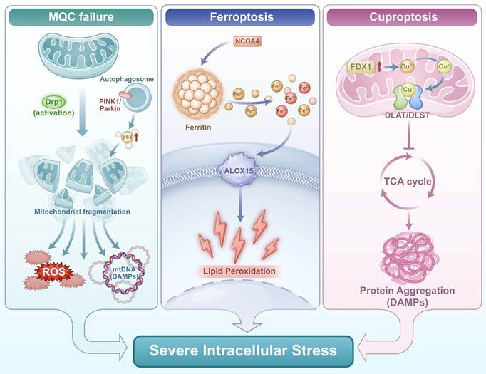
A tight bidirectional dialogue exists between metal homeostasis and MQC. On one hand, mitophagy is crucial for clearing excess metals, particularly iron; dysfunctional mitophagy leads to pathological metal retention within mitochondria[61]. On the other hand, ROS from iron overload damages mitochondrial DNA and promotes fission, while copper toxicity directly damages mitochondrial proteins, triggering compensatory (often overwhelmed) mitophagy. Together, MQC failure, iron-induced lipid peroxidation, and copper-induced proteotoxicity create an overwhelmingly hostile intracellular environment. We postulate that this metabolic crisis constitutes the critical priming event (Hit 1) for death network activation. The sustained cytokine storm (e.g., hyperactive IL-6/JAK/STAT3 signaling) amplifies this primed state by exacerbating mitochondrial ROS production and NLRP3 inflammasome activity. A subsequent triggering event — sensing of cytosolic nucleic acids by ZBP1 — pushes stressed kinase networks past their tipping point, initiating PANoptosis execution (Figure 1).
Convergence of MQC failure and metal dysregulation on severe intracellular stress. During sepsis, mitochondrial quality control (MQC) collapse triggers excessive Drp1-mediated mitochondrial fragmentation and impaired PINK1/Parkin-dependent mitophagy, leading to ROS overproduction and mtDNA (DAMP) release. Concurrently, NCOA4-driven ferritinophagy liberates free Fe²⁺, fueling iron-dependent lipid peroxidation (ferroptosis), while FDX1-mediated reduction of Cu²⁺ to Cu⁺ promotes copper-induced aggregation of lipoylated TCA cycle proteins (cuproptosis). The synergistic stress from these three pathways converges to generate a hostile intracellular environment, serving as the critical priming platform (Hit 1) for the cell death network in sepsis.
3.4 Molecular Mediators Linking MQC-Metal Stress to PANoptosome Activation
The transition from cellular stress (Hit 1) to death execution (Hit 2) is mediated by specific molecular signals that physically interact with and activate PANoptosome pattern recognition receptors. We propose the following mediators as key nodes connecting the priming and triggering phases:
(i) Oxidized mtDNA fragments: Mitochondrial dysfunction and iron-induced ROS lead to oxidative damage of mtDNA. Oxidized mtDNA fragments released into the cytosol serve as DAMPs that activate the cGAS-STING pathway, which in turn promotes ZBP1-PANoptosome assembly[27].
(ii) Lipid peroxidation-derived electrophiles: Products of ferroptotic lipid peroxidation, such as 4-HNE and malondialdehyde, can directly modify and activate NLRP3 inflammasome components, lowering the threshold for caspase-1 activation and GSDMD cleavage. ICU patients with simultaneous pyroptosis- and ferroptosis-positive signatures — defined by elevated IL-1Ra, IL-18, GDF15 (pyroptosis) and MDA, catalytic iron (ferroptosis) — have the lowest survival probability[62], providing clinical evidence for this crosstalk.
(iii) Z-RNA species: Ribosomal dysfunction and impaired ribophagy lead to accumulation of aberrant ribosomal RNA species with Z-conformation (Z-RNA), which are directly sensed by ZBP1, nucleating PANoptosome formation. NUFIP1-mediated ribophagy effectively clears aberrant ribosomes, thereby suppressing ZBP1-driven PANoptosome formation[27].
(iv) Mitochondrial ROS: ROS generated from dysfunctional mitochondria oxidize critical cysteine residues on RIPK1 and other PANoptosome components, promoting their activation and oligomerization[63].
(v) Itaconate: A landmark 2026 study[64] demonstrated that pathophysiologically relevant high concentrations of itaconate covalently alkylate the absent in melanoma 2 (AIM2) protein at the cysteine 113 (C113) residue. Itaconate-mediated C113 alkylation structurally stabilizes the AIM2 protein and triggers a conformational change, enabling it to drive ASC oligomerization, PANoptosome assembly, and subsequent macrophage PANoptosis. In vivo models further corroborated the pathogenic contribution of this axis to systemic sepsis. The AIM2 C113A mutation completely abolishes itaconate-induced AIM2 stabilization and PANoptosis. This reveals a novel pro-inflammatory mechanism of itaconate — an immunometabolite traditionally considered anti-inflammatory — via the post-translational modification of AIM2. This suggests that metabolic intermediates themselves may serve as PANoptosis triggers, and that the itaconate-AIM2 alkylation axis provides crucial mechanistic insights into macrophage depletion and systemic inflammation, highlighting a potential therapeutic target for severe sepsis.
These mediators collectively provide the molecular bridge between the metabolic priming environment and the nucleic acid-sensing triggering machinery. Their relative contributions may vary across cell types and sepsis phases, a question that warrants systematic investigation.
4. Metabolic Cell Death Programs: From Individual Pathways to the PANoptosis Hub
4.1 Ferroptosis: Iron-Dependent Lipid Peroxidation Amplification Loop
The core defense axis against ferroptosis is the System Xc⁻-GSH-GPX4 pathway. System Xc⁻ imports cystine for glutathione (GSH) synthesis; glutathione peroxidase 4 (GPX4) uses GSH to reduce phospholipid hydroperoxides[65]. In sepsis, inflammatory cytokines suppress SLC7A11, metabolic derangements limit GSH regeneration, and ROS inactivate GPX4. Acyl-CoA synthetase long-chain family member 4 (ACSL4) and lysophosphatidylcholine acyltransferase 3 (LPCAT3) incorporate PUFAs into membranes, shaping a ferroptosis-sensitive lipidome[66]. Alternative defenses (FSP1-CoQ10, GCH1-BH4) also exist[67-69]. Lipid peroxidation not only destroys membrane integrity but also primes cells for broader death networks.
4.2 Cuproptosis: Mitochondrial Protein Aggregation Catastrophe
As introduced in Section 3.2, cuproptosis is characterized by FDX1-dependent aggregation of lipoylated TCA proteins (DLAT, DLST) and Fe-S cluster disruption. It is independent of other death pathways. In sepsis, the heart and kidney are particularly vulnerable[52,55]. For memory T cells, copper-induced proteotoxic stress may cause metabolic paralysis — a hypothesis that remains to be tested.
4.3 Disulfidptosis and Parthanatos: Complementary Metabolic Death Routes
Two additional metabolic death pathways have been proposed in sepsis[70,71]. Disulfidptosis is triggered by glucose deprivation-induced NADPH depletion, leading to abnormal disulfide crosslinking of actin cytoskeleton proteins[70]. Parthanatos is initiated by severe DNA damage: hyperactivated poly (ADP-ribose) polymerase 1 (PARP1) consumes NAD⁺, depletes ATP, and triggers AIF release, causing large-scale DNA fragmentation[71]. While conceptually attractive given sepsis-induced metabolic disturbances and ischemia-reperfusion injury, direct in vivo evidence for these pathways in sepsis remains absent; they are currently classified as speculative mechanisms requiring dedicated investigation.
4.4 Pyroptosis: The Inflammatory Amplifier
Pyroptosis is tightly coupled to cellular metabolism. Inflammasome activation (e.g., NLRP3) triggers caspase-1, which cleaves gasdermin D (GSDMD); the N-terminal fragment forms membrane pores, releasing IL-1β and IL-18 and causing osmotic lysis[18]. NLRP3 activation is closely associated with a shift to aerobic glycolysis and relies heavily on mitochondrial ROS and released DAMPs (e.g., mtDNA)[72,73]. Pyroptosis and ferroptosis engage in feed-forward crosstalk: pyroptotic cells release factors that exacerbate oxidative stress, promoting ferroptosis, and lipid peroxidation products (e.g., 4-HNE) can directly activate the NLRP3 inflammasome, creating a self-amplifying inflammatory loop[74,75]. This synergy is particularly evident in sepsis-induced ARDS and acute kidney injury. In sepsis-associated encephalopathy (SAE), microglial pyroptosis has been identified as a key pathogenic mechanism. Wang[76] et al. demonstrated that activation of the α7 nicotinic acetylcholine receptor (α7 nAChR) reduces pyroptosis by enhancing microglial mitophagy, thereby mitigating cognitive deficits in mice with SAE. Similarly, pharmacological targeting of macrophage pyroptosis has emerged as a promising strategy in sepsis-induced ALI. Liu[77] et al. showed that BRD3308 suppresses macrophage oxidative stress and pyroptosis via upregulating H3K27 acetylation, thereby attenuating sepsis-induced lung injury.
Limitations of current biomarkers for distinguishing cell death modalities in vivo. A major challenge in validating the pyroptosis-ferroptosis crosstalk in sepsis is the lack of definitive biomarkers to distinguish these death pathways in tissue homogenates. Reliance on single markers — such as 4-hydroxynonenal (4-HNE) or malondialdehyde (MDA) for ferroptosis, or cleaved GSDMD for pyroptosis — is fraught with limitations: (i) lipid peroxidation products like 4-HNE and MDA are not specific to ferroptosis and can arise from general oxidative stress; (ii) cleaved GSDMD in tissue lysates may derive from non-pyroptotic caspase activation; and (iii) these markers do not provide spatial information about whether both death programs occur in the same cell or microenvironment. Notably, ICU patients with simultaneous pyroptosis- and ferroptosis-positive signatures — defined by elevated IL-1Ra, IL-18, GDF15 (pyroptosis) and MDA, catalytic iron (ferroptosis) — have the lowest survival probability[62], suggesting that combined biomarker panels, rather than single markers, offer superior prognostic value.
Recent advances in pyroptosis biomarkers for sepsis. Despite these limitations, significant progress has been made in developing more specific pyroptosis biomarkers. A recent study demonstrated that plasma Gasdermin D C-terminal fragment (GSDMD-CT) levels were significantly elevated in sepsis patients (23.02 pg/mL) compared to SIRS patients (16.52 pg/mL) and healthy controls (7.02 pg/mL), with an AUC of 0.881 (95% CI: 0.833-0.929), sensitivity of 85.0%, and specificity of 84.5% for sepsis diagnosis[78]. Another study reported an AUC of 0.870 with 91.0% sensitivity for GSDMD-CT in distinguishing sepsis from healthy controls[79]. These findings demonstrate that GSDMD-CT exhibits significant clinical value and holds great potential as a biomarker. These advances suggest that next-generation pyroptosis biomarkers — particularly those targeting specific GSDMD cleavage fragments — may overcome the limitations of traditional single-marker approaches and enable more precise endotyping of septic patients. To definitively establish pyroptosis-ferroptosis crosstalk in sepsis, future studies should employ multiplex immunohistochemistry or imaging mass cytometry to co-localize ferroptosis markers (e.g., ACSL4, 4-HNE-modified proteins) and pyroptosis markers (e.g., GSDMD N-terminus, NLRP3) at the single-cell level within the same tissue section. Single-cell RNA-seq or spatial transcriptomics could further resolve whether individual cells co-express signatures of both death pathways.
4.5 PANoptosis: A Master Hub Integrating Metabolic Priming and Nucleic Acid Sensing in Immune Exhaustion
4.5.1 The PANoptosome as an Integrated Death Platform
PANoptosis represents a conceptual leap in understanding sepsis-induced cell death. It is a coordinated, pro-inflammatory cell death pathway driven by a multifaceted molecular complex — the PANoptosome — which simultaneously integrates and executes pyroptosis, apoptosis, and necroptosis[19-21]. We propose a “Two-Hit” model:
Hit 1 (priming): MQC failure and iron/copper dyshomeostasis generate ROS and mtDNA leakage, lowering the cell's bioenergetic threshold for death network activation without committing it to execution. Critically, this primed state is not yet lethal on its own.
Hit 2 (triggering): Aberrant sensing of cytosolic nucleic acids by ZBP1, amplified by interferon signaling, nucleates PANoptosome assembly.
Within this framework, hyperactive IL-6/JAK/STAT3 signaling may synergize with intracellular metabolic stress not by directly activating RIPK1, but by creating a permissive inflammatory milieu that amplifies mitochondrial ROS production and NLRP3 inflammasome priming. Metabolic stress—particularly NAD⁺ depletion resulting from PARP1 hyperactivation—may indirectly lower the threshold for PANoptosome assembly by compromising the activity of key deubiquitinating enzymes that restrain RIPK1. The precise biochemical cascade linking NAD⁺ depletion to PANoptosome stabilization is detailed in Section 4.5.2. These hypotheses warrant direct biochemical validation in primary septic T cells.
4.5.2 Direct Evidence and Remaining Hypotheses
A recent study has provided direct evidence (Level A) of CD4⁺ T cell PANoptosis in sepsis, demonstrating that ZBP1 senses metabolic stress and assembles the PANoptosome, while NUFIP1-mediated ribophagy clears damaged ribosomes to restrain this process via cGAS-STING[27]. This confirms that PANoptosis is operative in septic T cells. Critically, however, the mechanistic connection between IL-6/JAK/STAT3 signaling and RIPK1-dependent PANoptosome assembly in CD4⁺ T cells remain a hypothesis (Level D) that has not been directly tested in primary septic T cells. While Kang[25] et al. provided evidence that IL-6 activates JAKs/STAT3 promoting RIPK1 phosphorylation and PANoptosome formation in CD4⁺ T cells from sepsis patients, this finding represents a single study requiring independent replication. Moreover, IL-6 blockade has yielded divergent outcomes across sepsis models[26], and the biochemical details of how STAT3 phosphorylation directly or indirectly modulates RIPK1 activity in the T cell context have not been elucidated. We therefore explicitly classify the IL-6/JAK/STAT3-RIPK1-PANoptosome axis in CD4⁺ T cells as a testable hypothesis requiring dedicated investigation using T cell-specific conditional knockouts and biochemical validation in primary cells, rather than an established mechanism.
Mechanistically, multiple layers of regulation link metabolic stress to PANoptosome assembly. First, ROS and lipid peroxides from MQC failure and ferroptosis may promote RIPK1 activation through cysteine oxidation or other post-translational modifications[63], though these have not been validated on specific residues in septic T cells. Second, NAD⁺ depletion—driven by PARP1 hyperactivation in the setting of severe DNA damage—reduces SIRT1 activity, an NAD⁺-dependent deacetylase[80]. Given that deubiquitinating enzymes such as CYLD and A20 are known to restrain RIPK1 activation by removing K63-linked polyubiquitin chains[81], it is hypothesized that NAD⁺ depletion may compromise deubiquitinase function and thereby lower the threshold for PANoptosome assembly. This indirect regulatory cascade (NAD⁺ depletion → SIRT1 inactivation → impaired deubiquitinase function → RIPK1 hyperubiquitination → PANoptosome stabilization) provides a testable biochemical hypothesis linking metabolic stress to PANoptosis execution, but requires direct validation in primary septic T cells. Notably, NAD⁺ precursor supplementation has been shown to increase NAD⁺ and SIRT1 levels, attenuate T cell exhaustion, and improve survival in preclinical sepsis models[80]. Third, recent evidence demonstrates that Zn²⁺ inhibits AIM2-mediated PANoptosome assembly via regulating mtDNA efflux[82]; fourth, inhibition of cIAP1/2 reduces RIPK1 phosphorylation in septic lung injury[83]; and fifth, itaconate covalently alkylates AIM2 at C113, driving macrophage PANoptosis during sepsis[64] — further expanding the repertoire of PANoptosome regulatory inputs that intersect with metabolic stress.
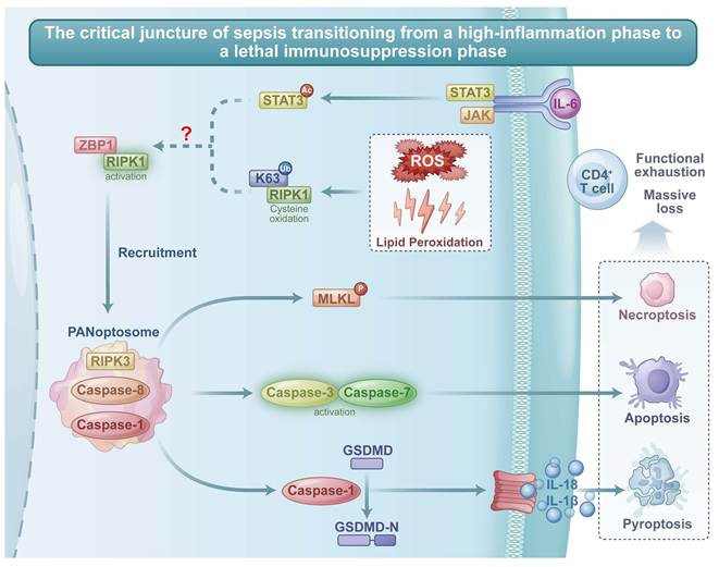
Thus, the direct demonstration of CD4⁺ T cell PANoptosis in sepsis has been achieved, but the IL-6/JAK/STAT3-RIPK1 linkage remains a hypothesis requiring separate testing using conditional knockout models (Figure 2).
The PANoptosis hub: integration of extrinsic cytokine signaling and intrinsic metabolic stress driving CD4⁺ T cell exhaustion. The collapse of mitochondrial quality control (MQC) and iron/copper dyshomeostasis generates a state of severe intracellular metabolic and oxidative stress — the priming hit (Hit 1) — characterized by ROS overproduction, lipid peroxidation, and mitochondrial DNA (mtDNA) leakage. This primed state lowers the bioenergetic and redox threshold for cell death but does not commit the cell to execution. The critical triggering hit (Hit 2) is provided by the aberrant sensing of cytosolic nucleic acids (mtDNA, Z-RNA) by the master sensor ZBP1, amplified by interferon signaling. In this model, the sustained IL-6/JAK/STAT3 cytokine storm may function as a context-dependent amplifier rather than a universal direct trigger, but creates a permissive and amplifying inflammatory environment that exacerbates mitochondrial ROS production, primes the NLRP3 inflammasome, and may impair STAT3-mediated pro-survival signaling. However, the direct contribution of IL-6/JAK/STAT3 to RIPK1 activation and PANoptosome nucleation in CD4⁺ T cells remains a testable hypothesis (Level D evidence) that requires dedicated experimental validation. Activated ZBP1 and redox-modified RIPK1 nucleate the assembly of the PANoptosome. Solid lines (e.g., ZBP1→PANoptosome, PANoptosome→Caspase-1/3/8/MLKL, and downstream effector pathways) indicate pathways with direct experimental validation (Level A-B evidence). Dashed lines with question marks (?) (e.g., IL-6/JAK/STAT3→RIPK1 and ROS→RIPK1) indicate hypothetical or contentious connections (Level D evidence) that require further validation in primary septic CD4⁺ T cells. Through the adaptor proteins FADD and ASC, this multimolecular complex simultaneously recruits and activates RIPK3, Caspase-8, and Caspase-1. This platform coordinately executes pyroptosis (via Caspase-1-mediated GSDMD cleavage and IL-1β/IL-18 release), apoptosis (via Caspase-3/7 activation), and necroptosis (via MLKL phosphorylation). This integrated, multimodal death program results in the massive depletion and functional exhaustion of CD4⁺ T cells, marking the fatal transition from hyperinflammation to immunosuppression in sepsis.
5. Organ and Cell-Type Specificity of the Death Network
The metabolic death network exhibits marked heterogeneity across organs and immune cell subsets. Table 1 summarizes the dominant death programs, key mechanisms, and therapeutic implications for each major site.
Several key patterns emerge. First, the pyroptosis-ferroptosis cycle represents a common amplification loop in multiple organs, particularly the lung and kidney. Second, the role of cuproptosis is only beginning to be defined, with the strongest evidence currently in the heart. Third, the massive depletion of CD4⁺ T cells via PANoptosis represents a systemic event that may underpin the transition from hyperinflammation to immunosuppression — a hypothesis that warrants further investigation with T cell-specific genetic models.
6. Clinical Translation: From Network Understanding to Precision Intervention
6.1 Multidimensional Biomarker Panels for Endotyping
An ideal multidimensional biomarker panel for sepsis endotyping should encompass: (i) death program activity markers (plasma 4-HNE for ferroptosis, extracellular N-terminal GSDMD for pyroptosis, circulating PANoptosome components; GSDMD-CT as a novel plasma biomarker for pyroptosis[79]); (ii) MQC and mitochondrial function markers (plasma cell-free mtDNA); (iii) immunometabolic signatures (single-cell metabolic flux, CD4⁺ T cell count with programmed cell death protein 1 (PD-1)/T-cell immunoglobulin and mucin domain-containing protein 3 (TIM-3); combined pyroptosis/ferroptosis biomarker panels — IL-1Ra, IL-18, GDF15, MDA, catalytic iron — have been shown to predict survival in ICU patients[62]); and (iv) traditional inflammatory markers (PCT, CRP, IL-6). The identification of reliable sepsis biomarkers is further complicated in specific patient populations. In burn injury, for instance, the systemic inflammatory response triggered by the thermal insult itself confounds conventional biomarker interpretation. Palmieri[84] et al. provided an updated overview of sepsis biomarkers specifically in this context, underscoring the need for population-adapted biomarker panels.
Integration of these data streams using machine learning can stratify patients into mechanistically defined endotypes (e.g., “Hyperinflammatory-Pyroptosis-Dominant,” “Metabolic Collapse-Ferroptosis/Cuproptosis-Dominant,” or “Immunoparalytic-PANoptosis-Dominant”) to guide trial design and personalized treatment[85,86].
6.2 Targeted Therapeutic Strategies
Biological risks of systemic cell death inhibition. Before discussing specific therapeutic strategies, it is essential to acknowledge the fundamental biological risks associated with systemically inhibiting cell death pathways in the context of active infection. First, broad inhibition of necroptosis or pyroptosis during active bacterial infection can impair pathogen clearance, as these death pathways are critical for eliminating infected cells and exposing intracellular bacteria to immune surveillance[18,19]. Second, indiscriminate copper or iron chelation poses risks of systemic metabolic toxicity, including copper deficiency-induced neurological and hematopoietic toxicity, and worsening anemia in patients already suffering from sepsis-associated anemia[60]. Third, many cell death inhibitors have off-target effects that may affect unrelated signaling pathways or cause unintended immunosuppression. Fourth, the timing of intervention is critical — inhibiting cell death too early may impair the host's ability to control the initial infection, while inhibiting too late may allow irreversible organ damage to occur. These risks underscore the urgent need for targeted delivery strategies (e.g., cell-specific nanocarriers, organ-selective formulations) that concentrate therapeutic agents at sites of pathological cell death while sparing essential immune functions. The development of such delivery systems represents a critical priority for translating cell death-targeting therapies from bench to bedside.
The imperative for targeted delivery. The risks outlined above argue strongly for the development of cell- and organ-specific delivery strategies rather than systemic administration of cell death inhibitors. Nanoparticle-based delivery systems offer several advantages: (i) passive targeting to inflammatory tissues via enhanced permeability and retention (EPR) effect; (ii) active targeting using cell-surface markers (e.g., CD4 for T cells, CD68 for macrophages, ACE2 for lung epithelial cells); (iii) stimulus-responsive release triggered by the inflammatory microenvironment (e.g., ROS-sensitive or pH-sensitive nanocarriers); and (iv) reduced systemic exposure and off-target toxicity. Preclinical studies have begun to explore such approaches in sepsis models, but this remains a nascent field requiring substantial investment. Until such targeted delivery systems are developed and validated, the clinical translation of systemic cell death inhibitors will remain limited by unacceptable toxicity profiles.
The following therapeutic strategies are presented as hypothetical directions based on preclinical and inferential evidence; none have been validated in clinical sepsis trials and should not be construed as clinical recommendations. Table 2 provides a comprehensive list of agents and their clinical stages. Importantly, the majority of these strategies remain at preclinical stages (Evidence Level B-C), and their clinical translation is hindered by off-target effects, tissue-specific delivery challenges, and the risk of unintended immunosuppression or metabolic toxicity (e.g., copper deficiency, impaired mitochondrial function with excessive chelation).
Recent progress highlights several promising approaches: (i) ferroptosis-targeting agents such as iron chelators (deferoxamine, best suited for patients with biochemical evidence of iron overload) and GPX4 stabilizers; (ii) copper-targeting strategies, where tetrathiomolybdate is now justified for preclinical testing in organ-specific sepsis models— though caution is warranted given that immoderate copper chelation was detrimental to mitochondrial function and cell viability[60]; (iii) pyroptosis and PANoptosis network modulators, including GSDMD inhibitors (disulfiram[87], necrosulfonamide[88], AI-designed peptide SK56[89]), inflammasome inhibitors (MCC950[90], VX-765[91]), and RIPK1 inhibitors — including the FDA-approved anti-epileptic drug phensuximide, which inhibits RIPK1-dependent immunogenic cell death and protects against LPS- and TNF-induced systemic inflammation in sepsis models[92] — as a master regulatory node to simultaneously dampen multiple death pathways; and (iv) MQC modulators and metabolic support (Drp1 inhibitor Mdivi-1[93], mitophagy enhancers like urolithin A[94], biogenesis agonists, thiamine[95], NAD⁺ precursors[80]).
6.3 Dynamic, Phase-Specific Combination Regimens: Embracing Temporal Heterogeneity
The following combination strategies are presented as hypothetical future directions based on preclinical and inferential evidence; they have not been validated in clinical trials and should not be construed as clinical recommendations. Therapy must be adapted to the dominant pathophysiology at a given time. However, the temporal boundaries between sepsis phases are not rigid and exhibit substantial inter-patient heterogeneity. Clinical and immunological evidence demonstrates that sepsis-induced immunosuppression, lymphocyte apoptosis, and T-cell exhaustion frequently initiate within the first 24 to 48 hours, concurrent with the hyperinflammatory cytokine storm[5,7]. Therefore, the following time windows should be interpreted as general heuristic guides rather than fixed therapeutic calendars, and all treatment decisions should be guided by real-time biomarker monitoring rather than chronological time alone.
Early phase (typically 0-48 h, but may extend or contract depending on pathogen, host factors, and prior immune status): Characterized by hyperinflammation with cytokine storm. A hypothetical combination could include an IL-6/JAK/STAT3 modulator + a GSDMD inhibitor + broad-spectrum antibiotics. In patients with early-onset immunosuppression (biomarker-defined), immune-stimulatory approaches may need to be considered earlier than the traditional 7-day threshold.
Middle phase (typically days 3-7, but may overlap with early or late phases in individual patients): Therapy should be guided by the primary failing organ: for ARDS, consider an inhaled anti-pyroptosis agent plus an anti-ferroptosis compound; for SA-AKI, a renally targeted ferroptosis inhibitor. Biomarker panels[62] (IL-1Ra, IL-18, GDF15, MDA, catalytic iron) should be used to determine whether pyroptosis-dominant, ferroptosis-dominant, or mixed signatures predominate in the individual patient.
Late phase (typically beyond day 7, but may occur earlier in patients with rapid progression): The focus shifts to immune stimulation (e.g., anti-PD-1 trials[96], GM-CSF, thymosin α1) and tissue repair, provided that PANoptosis execution was limited during the transition. Crucially, the transition to this phase should be defined by immune monitoring (e.g., persistent lymphopenia, HLA-DR expression, PD-1/TIM-3 upregulation) rather than calendar time.
A critical caveat: The three-phase framework described above is a heuristic simplification. In clinical reality, hyperinflammation and immunosuppression frequently coexist, with some patients exhibiting predominantly hyperinflammatory phenotypes throughout their ICU course, others showing early and profound immunosuppression, and still others oscillating between phenotypes[5,86]. This inter-patient heterogeneity mandates a biomarker-guided, adaptive approach rather than a one-size-fits-all chronological protocol. Combination regimens should be guided by real-time biomarker monitoring to avoid unintended reactivation of inflammation or premature immune stimulation in the presence of ongoing hyperinflammation.
7. Conclusion and Future Perspectives
The MQC-Metal-Death Program triad provides a powerful and cohesive lens through which to understand sepsis. We propose a testable two-hit model: MQC failure and metal dyshomeostasis act as the catastrophic priming event (Hit 1), lowering the threshold for death network activation, while ZBP1-mediated sensing of cytosolic nucleic acids serves as the triggering event (Hit 2) that drives PANoptosome assembly. Direct experimental testing of this hypothesis in conditional knockout models and human tissues is urgently needed before clinical translation can be considered.
Future research should prioritize four directions. First, dynamic multi-omics mapping—integrating single-cell RNA/ATAC profiling, immune-repertoire assays, and spatial transcriptomics—to resolve monocyte and T cell states in septic tissues[97]. Second, mechanistic dissection using conditional knockouts (Ripk1, Zbp1, Gsdmd, Nufip1), complemented by spatial transcriptomics and single-cell metabolomics to capture the transient 'primed' state of T cells in vivo. Third, biomarker-guided adaptive trials leveraging combined pyroptosis/ferroptosis panels (IL-1Ra, IL-18, GDF15, MDA, catalytic iron) that predict ICU survival[62], alongside GSDMD-CT as a novel early sepsis biomarker[79]. Fourth, resilience strategies including early metabolic support (NAD⁺ precursors, nicotinamide riboside) and lymphocyte PANoptosis prevention via ribophagy enhancement.
Key experiments to test the two-hit model:
(i) Temporal dissection: Longitudinal sampling of septic CD4⁺ T cells (0-72h post-CLP) to correlate MQC/metal markers with PANoptosis activation. Prediction: PANoptosis markers peak after MQC and metal dysregulation reach threshold.
(ii) Genetic epistasis: T cell-specific knockouts of Hit 1 components (Drp1, ferritin H) should attenuate Hit 2-induced PANoptosis; Hit 2 knockouts (Zbp1) should block PANoptosis execution without affecting priming.
(iii) Rescue experiments: MQC-restoring interventions (mitophagy enhancers, nicotinamide riboside) or metal chelation should raise the threshold for ZBP1-induced PANoptosis, with minimal effect once PANoptosome assembly is initiated.
(iv) Human validation: Longitudinal analysis of CD4⁺ T cells from septic patients, correlating MQC/metal markers with PANoptosis markers and clinical outcomes, using validated biomarker panels as a starting point[62].
Ultimately, conquering sepsis requires abandoning the "one-size-fits-all" paradigm and embracing precision medicine grounded in deep, dynamic molecular phenotyping.
Acknowledgements
Funding
National Natural Science Foundation of China (No. 82202387).
Consent for publication
All authors consent to the publication of the article. (This review article does not contain any individual person's data in any form.).
Author contributions
The conceptualization and design of the study were provided by Xijie Dong. Chuntao Wang and Qiqi Wu initiated the study and prepared the initial manuscript draft. Chaoyao Hou and Zhanfei Li took part in drafting, revising or critically reviewing the article. All authors reviewed the manuscript. Chuntao Wang and Qiqi Wu contributed equally to this work.
Use of AI and AI-assisted technologies
During the preparation of this work, the authors used DeepSeek solely for English language polishing and grammar checking. DeepSeek was not used for scientific data analysis, interpretation, or generation of references. After using this tool, the authors reviewed and edited the content as needed and take full responsibility for the content of the publication. No other AI or AI-assisted technologies were used.
Competing Interests
The authors have declared that no competing interest exists.
References
1. Singer M, Deutschman CS, Seymour CW. et al. The Third International Consensus Definitions for Sepsis and Septic Shock (Sepsis-3). JAMA. 2016;315(8):801-810 doi:10.1001/jama.2016.0287
2. GBD 2021 Global Sepsis Collaborators. Global, regional, and national sepsis incidence and mortality, 1990-2021: a systematic analysis. Lancet Glob Health. 2025;13(12):e2013-e2026 doi:10.1016/S2214-109X(25)00356-0
3. Marshall JC. Why have clinical trials in sepsis failed? Trends Mol Med. 2014;20(4):195-203 doi:10.1016/j.molmed.2014.01.007
4. Tuttle K, McDonald M, Anderson E. Re-evaluating biologic pharmacotherapies that target the host response during sepsis. Int J Mol Sci. 2019;20(23):6049 doi:10.3390/ijms20236049
5. Hotchkiss RS, Monneret G, Payen D. Sepsis-induced immunosuppression: from cellular dysfunctions to immunotherapy. Nat Rev Immunol. 2013;13(12):862-874 doi:10.1038/nri3552
6. Long B, Gottlieb M. Emergency medicine updates: Management of sepsis and septic shock. Am J Emerg Med. 2025;90:179-191 doi:10.1016/j.ajem.2025.01.054
7. Delano MJ, Ward PA. The immune system's role in sepsis progression, resolution, and long-term outcome. Immunol Rev. 2016;274(1):330-353 doi:10.1111/imr.12499
8. Lelubre C, Vincent JL. Mechanisms and treatment of organ failure in sepsis. Nat Rev Nephrol. 2018;14(7):417-427 doi:10.1038/s41581-018-0005-7
9. Lira Chavez FM, Gartzke LP, van Beuningen FE. et al. Restoring the infected powerhouse: mitochondrial quality control in sepsis. Redox Biol. 2023;68:102968 doi:10.1016/j.redox.2023.102968
10. Liao X, Wang Y, Zhang Z, Qu X, Zhou G. Bidirectional Regulation of NLRP3 Inflammasome and Mitochondrial Quality Control in Sepsis: Mechanisms and Therapeutic Implications. Mediators Inflamm. 2026;2026:3168669 doi:10.1155/mi/3168669
11. Ganz T. Systemic iron homeostasis. Physiol Rev. 2013;93(4):1721-1741 doi:10.1152/physrev.00008.2013
12. Nemeth E, Ganz T. Hepcidin-Ferroportin Interaction Controls Systemic Iron Homeostasis. Int J Mol Sci. 2021;22(12):6493 doi:10.3390/ijms22126493
13. Tang D, Chen X, Kang R, Kroemer G. Ferroptosis: molecular mechanisms and health implications. Cell Res. 2021;31(2):107-125 doi:10.1038/s41422-020-00441-1
14. Stockwell BR, Friedmann Angeli JP, Bayir H. et al. Ferroptosis: A Regulated Cell Death Nexus Linking Metabolism, Redox Biology, and Disease. Cell. 2017;171(2):273-285 doi:10.1016/j.cell.2017.09.021
15. Tsvetkov P, Coy S, Petrova B. et al. Copper induces cell death by targeting lipoylated TCA cycle proteins. Science. 2022;375(6586):1254-1261 doi:10.1126/science.abf0529
16. Mao C, Wang M, Zhuang L, Gan B. Metabolic cell death in cancer: ferroptosis, cuproptosis, disulfidptosis, and beyond. Protein Cell. 2024;15(9):642-660 doi:10.1093/procel/pwae003
17. Yang L, Guttman L, Dawson VL, Dawson TM. Parthanatos: Mechanisms, modulation, and therapeutic prospects in neurodegenerative disease and stroke. Biochem Pharmacol. 2024;228:116174 doi:10.1016/j.bcp.2024.116174
18. Shi J, Gao W, Shao F. Pyroptosis: Gasdermin-Mediated Programmed Necrotic Cell Death. Trends Biochem Sci. 2017;42(4):245-254 doi:10.1016/j.tibs.2016.10.004
19. Malireddi RKS, Kesavardhana S, Kanneganti TD. ZBP1 and TAK1: Master Regulators of NLRP3 Inflammasome/Pyroptosis, Apoptosis, and Necroptosis (PAN-optosis). Front Cell Infect Microbiol. 2019;9:406 doi:10.3389/fcimb.2019.00406
20. Zheng M, Kanneganti TD. The regulation of the ZBP1-NLRP3 inflammasome and its implications in pyroptosis, apoptosis, and necroptosis (PANoptosis). Immunol Rev. 2020;297(1):26-38 doi:10.1111/imr.12909
21. Samir P, Malireddi RKS, Kanneganti TD. The PANoptosome: A Deadly Protein Complex Driving Pyroptosis, Apoptosis, and Necroptosis (PANoptosis). Front Cell Infect Microbiol. 2020;10:238 doi:10.3389/fcimb.2020.00238
22. Rose-John S, Winthrop K, Calabrese L. The role of IL-6 in host defence against infections: immunobiology and clinical implications. Nat Rev Rheumatol. 2017;13(7):399-409 doi:10.1038/nrrheum.2017.83
23. Hirano T. IL-6 in inflammation, autoimmunity and cancer. Int Immunol. 2021;33(3):127-148 doi:10.1093/intimm/dxaa078
24. Hunter CA, Jones SA. IL-6 as a keystone cytokine in health and disease. Nat Immunol. 2015;16(5):448-457 doi:10.1038/ni.3153
25. Kang Z, Hou C, Qi S, Li Z, Bai X, Dong X. Interleukin-6-Associated Pyroptosis, Apoptosis, and Necroptosis via JAKs/STAT3-RIPK1 Axis: A Potential Mechanism for CD4+ T-Cell Depletion in Bacterial Sepsis. Crit Care Med. 2026;54(6):1365-1376 doi:10.1097/CCM.0000000000007088
26. Lee SI, Kim NY, Chung C. et al. IL-6 and PD-1 antibody blockade combination therapy regulate inflammation and T lymphocyte apoptosis in murine model of sepsis. BMC Immunol. 2025;26(1):3 doi:10.1186/s12865-024-00679-z
27. Zhao P, Li J, He P. et al. NUFIP1-Mediated Ribophagy Alleviates PANoptosis of CD4+ T Lymphocytes in Sepsis via the cGAS-STING Pathway. Research (Wash D C). 2025;8:0895 doi:10.34133/research.0895
28. Chen W, Zhao H, Li Y. Mitochondrial dynamics in health and disease: mechanisms and potential targets. Signal Transduct Target Ther. 2023;8(1):333 doi:10.1038/s41392-023-01547-9
29. Westermann B. Mitochondrial fusion and fission in cell life and death. Nat Rev Mol Cell Biol. 2010;11(12):872-884 doi:10.1038/nrm3013
30. Adebayo M, Singh S, Singh AP, Dasgupta S. Mitochondrial fusion and fission: The fine-tune balance for cellular homeostasis. FASEB J. 2021;35(6):e21620 doi:10.1096/fj.202100067R
31. Xiong C, Guan F, Feng J. et al. Prolonged sepsis triggers abnormal mitochondrial dynamics in the limb muscles and diaphragm. Mol Cell Biochem. 2025;480(11):5721-5739 doi:10.1007/s11010-025-05338-4
32. Hu D, Sheeja Prabhakaran H, Zhang YY, Luo G, He W, Liou YC. Mitochondrial dysfunction in sepsis: mechanisms and therapeutic perspectives. Crit Care. 2024;28(1):292 doi:10.1186/s13054-024-05069-w
33. Marchi S, Guilbaud E, Tait SWG, Yamazaki T, Galluzzi L. Mitochondrial control of inflammation. Nat Rev Immunol. 2023;23(3):159-173 doi:10.1038/s41577-022-00760-x
34. Wu F, Zhang YT, Teng F, Li HH, Guo SB. S100a8/a9 contributes to sepsis-induced cardiomyopathy by activating ERK1/2-Drp1-mediated mitochondrial fission and respiratory dysfunction. Int Immunopharmacol. 2023;115:109716 doi:10.1016/j.intimp.2023.109716
35. Riley JS, Tait SW. Mitochondrial DNA in inflammation and immunity. EMBO Rep. 2020;21(4):e49799 doi:10.15252/embr.201949799
36. Oka T, Hikoso S, Yamaguchi O. et al. Mitochondrial DNA that escapes from autophagy causes inflammation and heart failure. Nature. 2012;485(7397):251-255 doi:10.1038/nature10992
37. Pickles S, Vigié P, Youle RJ. Mitophagy and Quality Control Mechanisms in Mitochondrial Maintenance. Curr Biol. 2018;28(4):R170-R185 doi:10.1016/j.cub.2018.01.004
38. Ni R, Ji XY, Cao T. et al. Nicotinamide mononucleotide protects septic hearts in mice via preventing cyclophilin F modification and lysosomal dysfunction. Acta Pharmacol Sin. 2025;46(4):976-988 doi:10.1038/s41401-024-01424-3
39. Lee J, Giordano S, Zhang J. Autophagy, mitochondria and oxidative stress: cross-talk and redox signalling. Biochem J. 2012;441(2):523-540 doi:10.1042/BJ20111451
40. Mancias JD, Wang X, Gygi SP, Harper JW, Kimmelman AC. Quantitative proteomics identifies NCOA4 as the cargo receptor mediating ferritinophagy. Nature. 2014;509(7498):105-109 doi:10.1038/nature13148
41. Popov LD. Mitochondrial biogenesis: An update. J Cell Mol Med. 2020;24(9):4892-4899 doi:10.1111/jcmm.15194
42. Yuan L, Li J, Yin L. et al. 5'tiRNA-33-CysACA-1 promotes septic cardiomyopathy by targeting PGC-1α-mediated mitochondrial biogenesis. Int J Biochem Cell Biol. 2025;179:106714 doi:10.1016/j.biocel.2024.106714
43. Cilleros-Holgado P, Gómez-Fernández D, Piñero-Pérez R. et al. Mitochondrial Quality Control via Mitochondrial Unfolded Protein Response (mtUPR) in Ageing and Neurodegenerative Diseases. Biomolecules. 2023;13(12):1789 doi:10.3390/biom13121789
44. Lu Q, Sun X, Yegambaram M. et al. Nitration-mediated activation of the small GTPase RhoA stimulates cellular glycolysis through enhanced mitochondrial fission. J Biol Chem. 2023;299(4):103067 doi:10.1016/j.jbc.2023.103067
45. Gonsales Spindola D, Bahr A, Clark S. et al. Endothelial cells retain inflammatory memory through chromatin remodeling in a two-hit model of infection-induced inflammation. Am J Physiol Cell Physiol. 2025;329(6):C1994-C2012 doi:10.1152/ajpcell.00567.2025
46. Bastarache JA, Smith K, Jesse JJ. et al. A two-hit model of sepsis plus hyperoxia causes lung permeability and inflammation. Am J Physiol Lung Cell Mol Physiol. 2022;322(2):L273-L282 doi:10.1152/ajplung.00227.2021
47. Ni S, Yuan Y, Kuang Y, Li X. Iron Metabolism and Immune Regulation. Front Immunol. 2022;13:816282 doi:10.3389/fimmu.2022.816282
48. Kagan VE, Mao G, Qu F. et al. Oxidized arachidonic and adrenic PEs navigate cells to ferroptosis. Nat Chem Biol. 2017;13(1):81-90 doi:10.1038/nchembio.2238
49. Guo M, Xiong M, Shi J, Peng Q, Lin S, Xu F. IL-27 Aggravates Sepsis-Induced ARDS by Driving Macrophage Ferroptosis Through Activation of NCOA4-Mediated Ferritinophagy. J Inflamm Res. 2026;19:575933 doi:10.2147/JIR.S575933
50. Zhang P, Liu W, Wang S, Wang Y, Han H. Ferroptosis and Its Role in the Treatment of Sepsis-Related Organ Injury: Mechanisms and Potential Therapeutic Approaches. Infect Drug Resist. 2024;17:5715-5727 doi:10.2147/IDR.S496568
51. Xu W, Wu Y, Wang S. et al. Melatonin alleviates septic ARDS by inhibiting NCOA4-mediated ferritinophagy in alveolar macrophages. Cell Death Discov. 2024;10(1):253 doi:10.1038/s41420-024-01991-8
52. Yan J, Li Z, Li Y, Zhang Y. Sepsis induced cardiotoxicity by promoting cardiomyocyte cuproptosis. Biochem Biophys Res Commun. 2024;690:149245 doi:10.1016/j.bbrc.2023.149245
53. Pang G, Hu K, Ji J. et al. Investigating hub genes in the relationship between septic cardiomyopathy and cuproptosis and potential Chinese herbal drug candidates with bioinformatic tools. Minerva Cardiol Angiol. 2024;72(5):453-464 doi:10.23736/S2724-5683.23.06476-1
54. Siang W, Guoyun W, Yan F, Wenji L. Mettl1 mitigates sepsis-induced cardiomyopathy via inhibition of FDX1-dependent cuproptosis. Gene. 2026;978:149892 doi:10.1016/j.gene.2025.149892
55. Lv D, Yu Y. Copper-Induced Cell Death in Renal Diseases: Molecular Mechanisms and Therapeutic Implications. Drug Des Devel Ther. 2025;19:11849-11861 doi:10.2147/DDDT.S562664
56. Wang L, Jiang L, Wang J. et al. Reduced glutathione ameliorates sepsis-induced acute kidney injury in mouse model: involvement of cuproptosis. Ren Fail. 2025;47(1):2518227 doi:10.1080/0886022X.2025.2518227
57. Li Q, Wang T, Zhou Y, Shi J. Cuproptosis in lung cancer: mechanisms and therapeutic potential. Mol Cell Biochem. 2024;479(6):1487-1499 doi:10.1007/s11010-023-04815-y
58. Parsanathan R. Copper's dual role: unravelling the link between copper homeostasis, cuproptosis, and cardiovascular diseases. Hypertens Res. 2024;47(5):1440-1442 doi:10.1038/s41440-024-01636-4
59. Zhang J, Wu Y, Du Y. et al. Cuproptosis-Related Genes as Prognostic Biomarkers for Sepsis: Insights into Immune Function and Personalized Immunotherapy. J Inflamm Res. 2024;17:4229-4245 doi:10.2147/JIR.S461766
60. Yang J, Chen N, Zhao P. et al. Diminished expression of GLS in CD4+ T cells serves as a prognostic indicator associated with cuproptosis in septic patients. Shock. 2024;62(1):51-62 doi:10.1097/SHK.0000000000002370
61. Chen G, Kroemer G, Kepp O. Mitophagy: An Emerging Role in Aging and Age-Associated Diseases. Front Cell Dev Biol. 2020;8:200 doi:10.3389/fcell.2020.00200
62. Willemart C, Seurinck R, Stroobants T. et al. Potential of biomarker-based enrichment strategies to identify critically ill patients for emerging cell death interventions. Cell Death Differ. 2025;32(12):2284-2293 doi:10.1038/s41418-025-01545-0
63. Meng Y, Sandow JJ, Czabotar PE, Murphy JM. The regulation of necroptosis by post-translational modifications. Cell Death Differ. 2021;28(3):861-883 doi:10.1038/s41418-020-00722-7
64. Ma J, Chen Y, Wang P. et al. The alkylation of AIM2 by itaconate mediates macrophage PANoptosis during sepsis. Cell Mol Immunol. 2026;23(6):619-634 doi:10.1038/s41423-026-01414-x
65. Jiang X, Stockwell BR, Conrad M. Ferroptosis: mechanisms, biology and role in disease. Nat Rev Mol Cell Biol. 2021;22(4):266-282 doi:10.1038/s41580-020-00324-8
66. Doll S, Proneth B, Tyurina YY. et al. ACSL4 dictates ferroptosis sensitivity by shaping cellular lipid composition. Nat Chem Biol. 2017;13(1):91-98 doi:10.1038/nchembio.2239
67. Salem S, Han T, Alchoueiry M. et al. Targeting FSP1 to induce ferroptosis in chromophobe renal cell carcinoma. Oncogene. 2025;44(42):4075-4086 doi:10.1038/s41388-025-03562-2
68. Bersuker K, Hendricks JM, Li Z. et al. The CoQ oxidoreductase FSP1 acts parallel to GPX4 to inhibit ferroptosis. Nature. 2019;575(7784):688-692 doi:10.1038/s41586-019-1705-2
69. Kraft VAN, Bezjian CT, Pfeiffer S. et al. GTP Cyclohydrolase 1/Tetrahydrobiopterin Counteract Ferroptosis through Lipid Remodeling. ACS Cent Sci. 2020;6(1):41-53 doi:10.1021/acscentsci.9b01063
70. Zou W, Sai L, Sai W, Song L, Wang G. Diagnostic and prognostic value of disulfidptosis-related genes in sepsis. Infect Med (Beijing). 2024;3(4):100143 doi:10.1016/j.imj.2024.100143
71. Moura RD, Mattos PD, Valente PF, Hoch NC. Molecular mechanisms of cell death by parthanatos: more questions than answers. Genet Mol Biol. 2024 47Suppl 1(Suppl 1):e20230357. doi:10.1590/1678-4685-GMB-2023-0357
72. Luo L, Zhuang X, Fu L. et al. The role of the interplay between macrophage glycolytic reprogramming and NLRP3 inflammasome activation in acute lung injury/acute respiratory distress syndrome. Clin Transl Med. 2024;14(12):e70098 doi:10.1002/ctm2.70098
73. Zhong Z, Liang S, Sanchez-Lopez E. et al. New mitochondrial DNA synthesis enables NLRP3 inflammasome activation. Nature. 2018;560(7717):198-203 doi:10.1038/s41586-018-0372-z
74. Linkermann A, Stockwell BR, Krautwald S, Anders HJ. Regulated cell death and inflammation: an auto-amplification loop causes organ failure. Nat Rev Immunol. 2014;14(11):759-767 doi:10.1038/nri3743
75. Li J, Liu H, Shan Z, Zhong K, Liang Q. Molecular mechanisms and potential implications of ferroptosis, cuproptosis, and disulfidptosis in septic lung injury. Front Med (Lausanne). 2025;12:1615264 doi:10.3389/fmed.2025.1615264
76. Wang Q, Luo Q, Su Z. et al. Activation of the α7 nicotinic acetylcholine receptor mitigates cognitive deficits in mice with sepsis-associated encephalopathy by inhibiting microglial pyroptosis. World J Emerg Med. 2025;16(5):438-446 doi:10.5847/wjem.j.1920-8642.2025.099
77. Liu B, Li N, Liu Y. et al. BRD3308 suppresses macrophage oxidative stress and pyroptosis via upregulating acetylation of H3K27 in sepsis-induced acute lung injury. Burns Trauma. 2024;12:tkae033 doi:10.1093/burnst/tkae033
78. Lyu YX, Bi X, Liu Y, Cui SJ, Zhao LX, Gao G. et al. The efficacy of plasma gasdermin D C-terminal fragment in the early diagnosis of sepsis. J Pract Med. 2025;41(12):1899-1906 doi:10.3969/j.issn.1006-5725.2025.12.019
79. Li J, Luo Y, Liu Y. et al. A novel Gasdermin D C-terminal neo-epitope as a biomarker for pyroptosis in sepsis. Clin Biochem. 2026;141:111045 doi:10.1016/j.clinbiochem.2025.111045
80. Zhao GJ, Yang XY, Zhang C. et al. SUPPLEMENTATION WITH NICOTINAMIDE RIBOSIDE ATTENUATES T CELL EXHAUSTION AND IMPROVES SURVIVAL IN SEPSIS. Shock. 2023;60(2):238-247 doi:10.1097/SHK.0000000000002153
81. Liu X, Tang AL, Cheng J, Gao N, Zhang G, Xiao C. RIPK1 in the inflammatory response and sepsis: Recent advances, drug discovery and beyond. Front Immunol. 2023;14:1114103 Published 2023 Apr 5. doi:10.3389/fimmu.2023.1114103
82. Guo J, Meng S, Zhang J, Wang N, Guo F. Zn2+ regulates mitochondrial DNA efflux to inhibit AIM2-mediated ZBP1-PANoptosome pathway and alleviate septic myocardial injury. Chem Biol Interact. 2025;417:111525 doi:10.1016/j.cbi.2025.111525
83. Liu X, Li Y, Zhang W. et al. Inhibition of cIAP1/2 reduces RIPK1 phosphorylation in pulmonary endothelial cells and alleviate sepsis-induced lung injury and inflammatory response. Immunol Res. 2024;72(4):841-850 doi:10.1007/s12026-024-09491-8
84. Palmieri TL, Heard J. Biomarkers of sepsis in burn injury: an update. Burns Trauma. 2025;13:tkae080 doi:10.1093/burnst/tkae080
85. Komorowski M, Celi LA, Badawi O, Gordon AC, Faisal AA. The Artificial Intelligence Clinician learns optimal treatment strategies for sepsis in intensive care. Nat Med. 2018;24(11):1716-1720 doi:10.1038/s41591-018-0213-5
86. Seymour CW, Kennedy JN, Wang S. et al. Derivation, Validation, and Potential Treatment Implications of Novel Clinical Phenotypes for Sepsis. JAMA. 2019;321(20):2003-2017 doi:10.1001/jama.2019.5791
87. Hu JJ, Liu X, Xia S. et al. FDA-approved disulfiram inhibits pyroptosis by blocking gasdermin D pore formation. Nat Immunol. 2020;21(7):736-745 doi:10.1038/s41590-020-0669-6
88. Yang W, Tao K, Wang Y. et al. Necrosulfonamide ameliorates intestinal inflammation via inhibiting GSDMD-medicated pyroptosis and MLKL-mediated necroptosis. Biochem Pharmacol. 2022;206:115338 doi:10.1016/j.bcp.2022.115338
89. Sun J, Yang J, Tao J. et al. Delaying pyroptosis with an AI-screened gasdermin D pore blocker mitigates inflammatory response. Nat Immunol. 2025;26(10):1660-1672 doi:10.1038/s41590-025-02280-x
90. Zheng Y, Zhang X, Wang Z. et al. MCC950 as a promising candidate for blocking NLRP3 inflammasome activation: A review of preclinical research and future directions. Arch Pharm (Weinheim). 2024;357(11):e2400459 doi:10.1002/ardp.202400459
91. Wannamaker W, Davies R, Namchuk M. et al. (S)-1-((S)-2-{[1-(4-amino-3-chloro-phenyl)-methanoyl]-amino}-3,3-dimethyl-butanoyl)-pyrrolidine-2-carboxylic acid ((2R,3S)-2-ethoxy-5-oxo-tetrahydro-furan-3-yl)-amide (VX-765), an orally available selective interleukin (IL)-converting enzyme/caspase-1 inhibitor, exhibits potent anti-inflammatory activities by inhibiting the release of IL-1beta and IL-18. J Pharmacol Exp Ther. 2007;321(2):509-516 doi:10.1124/jpet.106.111344
92. Kim BJ, Hong SM, Noh HJ. et al. FDA-approved phensuximide inhibits RIPK1-dependent immunogenic cell death. Cell Death Dis. 2025;16(1):426 doi:10.1038/s41419-025-07754-2
93. Zhang X, Fan H, Su L, Wang Y, Chen G. Mdivi-1 Attenuates Sepsis-Associated Acute Lung Injury by Inhibiting M1 Alveolar Macrophage Polarization and Pyroptosis. Mediators Inflamm. 2025;2025:3675276 doi:10.1155/mi/3675276
94. Ryu D, Mouchiroud L, Andreux PA. et al. Urolithin A induces mitophagy and prolongs lifespan in C. elegans and increases muscle function in rodents. Nat Med. 2016;22(8):879-888 doi:10.1038/nm.4132
95. Pereira AG, Costa NA, de Moraes MB. et al. Thiamine supplementation in septic shock patients: still looking for the target population. Crit Care. 2024;28(1):226 doi:10.1186/s13054-024-05012-z
96. Hotchkiss RS, Colston E, Yende S. et al. Immune checkpoint inhibition in sepsis: a Phase 1b randomized study to evaluate the safety, tolerability, pharmacokinetics, and pharmacodynamics of nivolumab. Intensive Care Med. 2019;45(10):1360-1371 doi:10.1007/s00134-019-05704-z
97. Yang Y, Zhang Y, Wu J, Liu Y, Lei X. Decoding immune low-response states in sepsis: single-cell and 3D spatial transcriptomic insights into immunoparalysis. Front Immunol. 2025;16:1696914 doi:10.3389/fimmu.2025.1696914
Author contact
Corresponding author: Xijie Dong, Tel: +86-027-83665913, E-mail: dr.dongxijieco.