Impact Factor ISSN: 1449-1907




Int J Med Sci 2026; 23(11):3440-3452. doi:10.7150/ijms.135665 This issue Cite
Review
Reprogramming of Human Limbal Niche Progenitors
Xinghan Guo1,2, Wei Wang1,2, Allison Wilde3, Yingting Zhu3  , Guigang Li1,2
1. Department of Ophthalmology, Tongji Hospital, Tongji Medical College, Huazhong University of Science and Technology, Wuhan 430030, China.
2. Hubei Key Laboratory of Otolaryngologic and Ophthalmic Diseases, Wuhan, 430000, China.
3. BioTissue, Miami, FL, 33126 USA.
Received 2026-4-5; Accepted 2026-9-9; Published 2026-9-24
Abstract
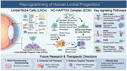
Limbal niche cells (LNCs) possess embryonic stem cell (ESC) characteristics, expressing ESC markers such as Sox2, Nanog, and Oct4; stromal characteristics, expressing neurocrest markers such as CD90; and have angiogenic potential expressing angiogenesis markers such as CD34. In addition, these cells can be induced into adipocytes, chondrocytes, and osteocytes, indicating that they are valuable adult stem cells. However, how these cells may be appropriately passaged and maintained, what signaling pathways are necessary for such regeneration, and how the cells can be used for the treatment of various diseases caused by stem cell deficiency, such as limbal stem cell deficiency, remain unclear. Herein, we summarize the most recent advances in limbal niche research, with emphasis on the regeneration of adult limbal progenitors and their mechanisms, to promote cell-based therapy for various eye diseases due to stem cell deficiency or malfunction. Such a strategy may also provide a platform for stem cell therapy for other similar human diseases worldwide.
Keywords: limbus, niche, stem cell, differentiation
Human Limbal Niche Cells
Limbal niche cells (LNCs) contain pigment cells (melanocytes) [1-3], neural crest cells, and vascular endothelial cells [4]. Although LNCs were suggested in those studies, their presence was later discovered and confirmed by our studies using collagenase [5, 6]. This enzymatic digestion technique resulted in the identification of LNCs as specific Vimentin (Vim)+ mesenchymal cells [5, 7]. Specifically, we reported that dispase digestion alone removes the sheets composed of limbal epithelia [8] but was unable to take away all limbal epithelial progenitors. In contrast, collagenase A digestion can remove all limbal progenitors and underlying pancytokeratin (PCK)-, p63α-, Vim+ stromal cells, which are small in size and express ESC markers such as Sox2 and CD34. Intriguingly, Bhartiya et al. also reported highly potent stem cells, small in size, expressing embryonic stem cell markers such as Nanog and Sox-2 [9]. In addition, human Limbal Niche Cells (hLNCs) have angiogenic potential, expressing angiogenesis markers, Fetal Liver Kinase-1 (Flk-1), CD34, CD31, and PDGFRβ [10].
The vascular niche contains basement membrane (BM), cellular components such as vascular endothelial cells (VECs) and pericytes [11]. The vascular BM contains laminin, collagen IV, and proteoglycans, with growth factors such as basic fibroblast growth factor (bFGF), epidermal growth factor (EGF), insulin-like growth factor (IGF), platelet-derived growth factor (PDGF), and transforming growth factor-β (TGF-β). Human SCs change from an osteoblastic to a vascular status to promote their replication and differentiation in the vascular BM [12, 13]. Human ESCs may remain undifferentiated when cultivated on top of Matrigel [14, 15]. BM promotes replication as well as differentiation in human stromal SCs in vitro [16, 17]. VECs can be supportive of bone marrow SCs [18], the central nervous system [19, 20], muscles [21], and pancreatic islets [22]. Their mechanisms involve physical contact and growth factors such as bFGF, Ang1, Bone Morphogenetic Protein 4 (BMP4), Desert Hedgehog Protein (DHH), Pigment Epithelium-Derived Factor (PEDF), NFC Data Exchange Format (NDEF), FGF4, and Stromal Cell-Derived Factor-1 (SDF-1) [12, 19, 20, 23]. Nerve growth factor (NGF) signaling may also promote the proliferation of limbal progenitors, increase the efficiency of colony formation, and maintain the stem cell phenotype [24].
MSCs within their Stem Cell Niche
Mesenchymal stem cells (MSCs) are located around perivascular areas of a number of tissues such as the bone marrow and the brain and spinal cord [25], the dental pulp [26], and skeletal muscle, pancreas, adipose tissue, and placenta [27]. Although the cell origin of MSCs is not very clear, accumulating evidence suggests that the progenitor cells are located in perivascular sites [28-30]. Interestingly, significantly different cell phenotypes are observed depending on cell location. For example, basal niche cells, in contrast to the cells around the rim, show distinct characteristics, suggesting that the fate of stem cells varies significantly depending on their position within or near the niche [31], likely due to differences in the microenvironment. Such a niche environment with more progenitors may be associated with the success rate of in vitro transplantation of epithelial stem cells because a higher number of autologous donor cells is closely associated with a higher transplantation success rate in the long term [32], and transplantation of limbal epithelial progenitors along with stromal cells is recommended for eye tissue repair and healing [33]. Currently, the prevailing approaches for restoring the microenvironment of the ocular surface are (1) cell-based therapies using progenitors capable of supporting the limbal epithelial progenitors and modulating inflammation by MSCs, (2) regeneration of the microenvironment using a bioactive matrix from decellularized tissues, such as amniotic membrane (AM), or synthetic molecules, and (3) promotion of certain signaling pathways using cytokines and growth factors [34-37].
HC-HA/PTX3 Complex
Human amniotic membrane (AM) may be applied at injury and surgical sites to inhibit inflammation, scarring (for reviews, see [38-40]), and regulate the niche of limbal epithelial progenitors [41-44]. Human AM may result in rapid regression of edema and inflammation of the corneal stroma [45-47]. Subsequently, our studies demonstrated that AM extract and HC-HA, where HC refers to the heavy chain from inter-α-inhibitor and HA refers to hyaluronan. Such HC-HA extracted from AM has functions against inflammation and scarring [48, 49]. Notably, HC-HA is more powerful against angiogenesis than HA by reducing migration, proliferation, differentiation, and viability without affecting Human Umbilical Vein Endothelial Cells (HUVECs) in culture [50]. We have shown that HC-HA can be reconstituted in vitro using purified components, e.g., HA, IαI, TNF-stimulated gene-6 (TSG-6), and TNF-stimulated gene 14 (TSG-14) (also known as Pentraxin 3 [PTX3]) [51]. For this purpose, others have added TSG-6 to create recombinant HC-HA (rcHC-HA) [52]. We, however, find that recombinant HC-HA thus created is different from that created when PTX3 is added first to immobilized HA. TSG-6, an important mediator of anti-inflammation and tissue remodeling [53, 54], is not detectable but can be quickly induced by IL-1 and TNF-α in many cell types [54-56].
HC-HA/PTX3 Supports Progenitor Status of Limbal Niche Cells
We have established a new approach for isolating and maintaining limbal epithelial progenitors and niche cells by digesting human limbus using collagenase [5]. Such LNCs, accompanied by limbal epithelial progenitors (LEPs), exhibit characteristics of being vimentin-positive, pancytokeratin-negative, and expressing SC markers, for example, Nanog, CD34, N-cadherin, Rex1, and SSEA4 [5, 57]. Such cells might be passaged on top of Matrigel, in a stem cell culture medium, to as many as 12 passages, exhibiting characteristics of MSCs and angiogenic potential [10]. When reseeded in three-dimensional (3D) cell culture matrix (Matrigel), these cells may revert to embryonic SC (ESC)-like cells. Based on the findings, we have established a 3D Matrigel model system in vitro, in which limbal epithelial progenitors (LEPs) may reunite with LNCs, resulting in sphere formation through activation of the SDF-1/CXCR4 (chemokine receptor type 4) cascade [7]. When LNCs are in constant contact with LEPs, better clonal growth is observed when cultured on top of 3T3 layers [5], which prevents LEPs from differentiating into corneal endothelial cells [57, 58]. Our studies demonstrated that activation of BMP and Wnt signaling pathways in both LNCs and LEPs, and also between LEPs and LNCs, resulted in LEPs' growth in the form of colonies. As a result, the spheres formed on the Matrigel inhibited corneal endothelial differentiation and promoted LEPs growth as colonies through the activation of the canonical Wnt pathway [59]. With HC-HA/PTX3 immobilized on the culture plates, sphere formation also suppressed corneal endothelial differentiation, promoted epithelial quiescence, and inhibited LEPs growth in colonies [60]. The results were linked to the inhibition of canonical Wnt and the activation of BMP and noncanonical Wnt pathways in both LNCs and LEPs. The BMP pathway from LNCs plays a critical role in the process, because inhibition of the BMP pathway in LEPs promotes cell cycle progression, inhibits self-renewal through Bmi-1, and increased LEPs growth in colonies. Therefore, HC-HA/PTX3 may activate the BMP pathway in both LNCs and LEPs, causing quiescence in vivo [61, 62] and ex vivo [63] while regulating the proliferation of LEPs, which might be a reliable candidate for the potential treatment of corneal blindness, for example, blindness caused by limbal stem cell deficiency (LSCD) [44, 64-66].
BMP-MET Initiates Adult Progenitor Reprogramming
Mesenchymal to epithelial transition (MET) occurs when tissues are being built or remodeled during embryogenesis, tissue repair, tissue restoration, fibrogenesis, and neoplasia. Unfortunately, it is still largely unknown how MET occurs. Recently, Takahashi was the first to show that somatic cells may be reprogrammed through Sox2, Krüppel-like Factor 4 (Klf4), Oct4, and MYC to induced pluripotent stem cells (iPSCs) [67]. This process of iPSC generation from adult mouse fibroblasts involves a change of cell shape from a spindle shape to tightly packed round clusters, resembling MET [67]. In the initial phase of reprogramming, it is reported that upregulation of E-cadherin and occludin [68-70] and downregulation of snail, a MET regulator [71], play a crucial role in achieving close cell-cell contact [70]. Ablation of endogenous E-cadherin reduces the reprogramming colony efficiency [68]. In vivo, knockout of E-cadherin leads to cellular decompaction during the mouse preimplantation process in early embryogenesis [72]. In our case, we did not detect any change in E-cadherin at the mRNA level in LNCs cultured on immobilized HC-HA/PTX3, indicating that factors other than E-cadherin are important during compaction. A previous report suggested that adult cellular reprogramming to iPSCs involves a mechanism in which reprogramming factors such as c-Myc, Oct4, and Sox2 inhibit Wnt and canonical TGF-β signaling [68]. Because Zeb siRNA combined with Y-27632, a small molecule that attenuates Rho-associated protein kinase (ROCK) proteins (ROCK1 and ROCK2), can completely reverse epithelial-to-mesenchymal transition (EMT) through eliminating contractile actomyosin bundles, reducing stromal mRNA and protein expression, and restoring E-cadherin and Ksp-cadherin that maintain healthy tubular epithelial cells [73], and Y27632 inhibits proliferation and EMT as we reported recently [74, 75], we speculate that inhibition of Rho-ROCK signaling is also important for MET, quiescence, and cell shape change. In addition, we have recently summarized how to avoid EMT for generating functional human corneal endothelia from their progenitors [76] and determined that progenitors are present in the trabecular meshwork [77].
Takahashi and Yamanaka et al. have characterized that reprogramming of adult cells into iPSCs may be achieved by the Sox2, Klf4, Oct4, and c-Myc (SKOM) method [67]. Three phases are involved in such reprogramming, i.e., the initiation, maturation, and stabilization phases (reviewed in [78]). The initial phase of reprogramming involves cell shape changes from a spindle shape to tightly packed round clusters, resembling MET [67]. In this phase, BMPs induce MET, causing significant shape changes; for example, the shape of fibroblasts may resemble that of epithelial cells [79, 80]. Blockade of BMPs impairs the initiation phase of the reprogramming process [70, 80]. The hallmark of MET change requires the downregulation of EMT regulators, such as snail1/2, slug, and Zeb1/2 [81], and the upregulation of E-cadherin, occludin, Cdh1, Crb3, EpCAM, and the epithelial-associated miRNA-200 family [68, 70, 82]. A recent study revealed that the miR-200 family promotes MET by suppressing Zeb2 and snail2. During these initial stages, several groups have demonstrated that cells acquire ESC-like proliferation, resistance to apoptosis, and changes in senescence cascade pathways [81, 83-85]. For instance, the BMP-SMAD-ID pathway inhibits cell senescence by inhibiting p16/INK4A [80]. In addition, a time-course study also disclosed that multiple markers, for instance, Thy1 (CD90), CD44, and ICAM1 (CD54), were lost while alkaline phosphatase and Tra-1-60 were gained. The epigenetic regulators Tet1, Tet2, and Tet3 are also required to activate the miR-200 family. Although the transition from the initiation phase to the next phase is not clearly defined, cells generally differentiate until the pluripotent genes are expressed. At this maturation phase, the detection of pluripotency-associated genes, including Fbxo15, Sall4, Oct4, and Nanog [70], can be used as a reliable assessment of successful reprogramming. It is suggested that adult cells may acquire pluripotent markers in a sequential way, in which markers such as Sox2 and CD31 are expressed during the stabilization phase. BMP signaling present in serum and feeders decreases iPSC generation and therefore must be downregulated or eliminated at the stabilization stage [80], and interestingly, Wnt activation is necessary in the late-stage reprogramming [86]. In the stabilization phase, cells are self-renewed, endogenous pluripotent genes are reactivated, the inactive X chromosome is restarted, and telomeres are lengthened, which restore normal cellular and tissue functions [79]. It remains unclear whether continuous BMP signaling during the stabilization phase promotes proper lineage differentiation (lineage-specific regeneration).
Studies have demonstrated that SCs in the niche compartments are regulated by several pathways, including BMP, Wnt, and Notch [87-90]. Among those signaling pathways, BMP signaling can be blocked by noggin [90-92], gremlin1 (GREM1), GREM, and chordin-like 1 (CHRDL1) [93, 94]. It is suggested that BMP signaling is closely mediated through BMP inhibitors, which prohibit BMPs from interacting with their receptors. These inhibitors may be classified into 3 main groups: (1) The BMP pro-region might fold back onto BMP proteins, act like a shield, thus blocking BMP interactions with their receptors; (2) Ligand inhibitors may bind to BMPs directly to form inactive complexes; and (3) Receptor inhibitors may interfere with BMP interactions with their receptors [95]. Chemicals such as sclerostin, follistatin, and noggin have been used as BMP inhibitors for BMP mechanistic studies [96-100]. Chordin and Connective Tissue Growth Factor (CTGF) are BMP antagonists that may act in the skeleton to regulate BMP availability [101]. In addition, other BMP antagonists, for example, Cerberus has a head-inducing ability in Xenopus but is not crucial for knockout mice [102, 103]; USAG-1 is a main regulatory protein that mediates tooth development and growth in a mouse model [104]; Coco is responsible for left-right symmetry [105]; and Prdc, Twisted, and Crossveinless-2 are associated with abnormal phenotypes, skeletal complications, and kidney and eye development [106]. Furthermore, BMP receptor antagonists include BMP-3, which is produced by osteoblasts, acts within bone tissue, and inhibits BMP activation via the type II activin receptor, thereby retarding bone growth [107, 108].
Cytoskeleton Changes Mediated by Rho GTPases
Compelling evidence suggests that both actin cytoskeletons and microtubules can be directly regulated by the Rho GTPase family (Rac, RhoA, Cdc42). RhoA can target ROCK (Rho kinase), which facilitates several downstream pathways involving cytoskeletal changes. First, ROCK can directly regulate the phosphorylation/dephosphorylation of the regulatory light chain (RLC) through a calcium-activated enzyme called myosin light chain kinase (MLCK) or the regulatory subunit of myosin phosphatase (MYPT1), respectively. This event prompts the activation of non-muscle myosin IIA and myosin IIB, which drive contractility of the cell cortex, cell motility, and disassembly of the actin network effectively at the cellular level. Another ROCK target is activated via LIM kinase (LIMK), which phosphorylates and inhibits cofilin. Activation of LIMK is not uniquely controlled by ROCK; however, downstream of activation of Rac1 and PAK can also activate LIMK or Testis-Associated Actin Remodeling Kinase 2 (TESK2), through phosphorylation at Ser3, thus inactivating the actin depolymerization factor (ADF)/cofilin. Meanwhile, the Slingshot (SSH) phosphatase and the HAD-family phosphatase, Chronophin (CIN), are dedicated to dephosphorylating cofilin. Interestingly, counteractions between Rac1 and RhoA may lead to different cellular behaviors (cellular heterogeneity), that is, they may act on identical genes but behave quite differently from one another, as demonstrated during epidermal morphogenesis. This study revealed that Rac1 through β-PIX/PIX-1 and PAK1, and RhoA, via ROCK/LET-502, may coordinate with each other to regulate apical junctions and the formation of basolateral protrusions during epidermal morphogenesis. Cell shape changes may be accomplished by generating physical forces through the following reactions: First, F-actin and myosin II work together to produce a pulling force with another cellular component, the cell cortex, in which E-cadherin acts as a linker between the inner network and the outside environment [109]. F-actin, cellular fibers acting as an internal framework, work together with myosin, a cellular motor protein, to create cellular force and movement, with epithelial cells as an important linker of various organs such as skin [110-116]. Rho GTPases turn on cellular switches by binding to GTP, becoming active, traveling to the plasma membrane, and triggering downstream cascades to mediate the cytoskeleton [117]. Previous work has identified Guanine Nucleotide Exchange Factors (GEFs) as the activators of RhoA during development [112, 118, 119]. For example, RhoA GEF2 proteins activate the RhoA pathway, which induces cellular shape changes with tissue folding during fruit fly embryonic development [118, 120]. Apical constrictions push tall cells into a wedge-like state, that is, a flat layer becomes bent and folded [121, 122]. Transcription factors, such as Snail and Twist, may promote G protein-coupled receptor signaling to mediate apical accumulation of RhoGEF2 [120, 123-125]. Apical RhoGEF2 acts as a switch to squeeze the apex of a cell, causing cell shape change. Myosin acts as a muscle within cells to lead to such a cellular change [113-116]. Myosin also acts as a small motor to propel cells to change shape and move by repeated short pulses during terminal contractions [126]. ROCK is also involved in cell shape change and movement in repeated beats [115, 116]. Cellular pulses probably occur due to myosin motors turning on and off in cycles, during which ROCK activates myosin by mediating its phosphorylation and dephosphorylation [115, 116], and possibly F-actin assembly is also involved in the process [113]. Myosin may cause F-actin contraction that dictates cellular shape changes [113, 126] and might help mediate cell signals in a timely manner at the correct cellular locations [116, 127]. In addition, myosin activities alter over time, and RhoA helps interacting proteins to form certain patterns to mediate such a cascade [113]. During this process, radial cell polarity (RCP) occurs, in which ROCK is rearranged in a unique pattern in the apical cellular compartment but absent at adherent junctions [113]. RCP is dependent on Twist. Interestingly, RhoA stimulation is needed but sufficient by itself to promote cellular apical contraction and interaction of ROCK and myosin. RhoGEF2 triggering might occur ahead of myosin activation, indicating that RhoA plays a significant role in contractile pulses. Such apical constriction requires Cumberland GAP (C-GAP) to control the location and the timing of RhoA proteins' activation within cells [128]. In fact, C-GAP controls whether the cell shape change is transient or stable by acting as a one-way lock [128, 129]. In all, present evidence suggests that activation of RhoA alone cannot induce cellular constriction. In contrast, RhoA signaling regulated by its helper proteins, RhoGEF2 and C-GAP, is the main mechanism mediating apical contraction and tissue status, as well as mediating the GEF-to-GAP ratio for a one-way-lock constriction into correct tissue alignment [128, 129]. Rac and Rho GTPases can control cell motility by changes in GTP hydrolytic activities, which are involved in cytoskeleton rearrangements [130].
SDF-1/CXCR4 Signaling
In the stem cell-based repair, the SDF-1/CXCR4 pathway plays a significant role in guiding stem cells into wounded tissues across multiple organs [131-133]. Late SDF-1 expression could contribute significantly to a therapeutic approach for tissue regeneration [131, 134]. Acute injury may trigger SDF-1 release, which attracts natural tissue stem cells to the damaged sites for repair [135] and maintains target organ cells in a healthy state, for instance, protecting heart muscle cells [136]. In 2011, Xie et al. from our lab reported that after cell-cell junctions were dissociated with trypsin and EDTA, individual NC and SC could be reassembled, leading to sphere formation on 3D Matrigel with embryonic SC culture medium, and that the expansion of spheres was mediated via SDF-1 produced by limbal epithelial progenitors and CXCR4 produced by limbal mesenchymal progenitors [7]. CXCR4 attenuation via a CXCR4-neutralizing antibody or the CXCR4 inhibitor AMD3100 attenuated the reunion and produced small, separate spheres. The resultant epithelial spheres were significantly differentiated, with a loss of holoclones, indicating that NC association regulated via SDF-1 and CXCR4 is essential for limbal SC performance [7]. We have recently shown that HC-HA/PTX3 may rejuvenate aging eye cells through important eye NC self-renewal and repair by turning senescent cells into NC progenitors via the SDF-1/CXCR4 pathway, which then triggers BMP activation to enhance cell growth and tissue repair. We deduce that HC-HA/PTX3-mediated rejuvenation may serve as a new medical therapy in the field of ophthalmology [137] (also reviewed in [138]). We have indicated that HC-HA/PTX3 can inhibit α-SMA expression and nuclear pSMAD2/3 translocation in human corneal fibroblasts even with the addition of TGF-β1, and such inhibition is accompanied by a phenotypic reversal to younger keratocytes via the activation of BMP signaling [139]. Further studies suggest that such phenotypic reversal is caused by cell aggregation initiated through SDF-1/CXCR4 signaling, showing CXCR4 nuclear translocation and upregulation of CXCR4 mRNA and protein, which activates canonical BMP signaling [139].
This new approach of epithelial cell growth in spheres mixed with NCPs helps illustrate how limbal progenitors renew and select their fate inside the limbal niche environment [140]. Chemokine SDF-1 is part of a large family of small, secreted proteins that mediate white blood cell movement in immune defense [141]. CXCR4 and CXCR7, identified as G-protein-coupled receptors (GPCRs), are the primary receptors for SDF-1 signaling [142, 143]. However, CXCR4, but not CXCR7, regulates GPCR signaling [143]. Our previous report indicates that limbal stromal cells express abundant CXCR4 but not CXCR7. CXCR4 attenuation by the CXCR4 inhibitor AMD3100 interfered with their reunion and generated separate, smaller spheres, which displayed significantly increased corneal differentiation and a notable decrease in holoclones [7]. Moreover, IGF-1 modulates CXCR-4 in human lung microvascular endothelial cells, indicating that IGF-1 may be upstream of CXCR-4 [144]. SDF-1 may enhance HGF expression in MSCs, suggesting that HGF may be downstream of SDF-1 signaling [145]. FGF2 may inhibit SDF-1 post-transcriptionally in bone marrow stromal cells via FGFR1 IIIc, suggesting that FGF2 is upstream of SDF-1 [146]. SDF-1 is a JUN-mediated factor critical in epithelial and mesenchymal interactions by mediating the expression of colony-stimulating factor 2 (CSF2) and fibroblast growth factor 7 (FGF-7) [147].
Notch Signaling
During eye development, activation of Notch signaling has been shown to induce Pax6 and other eye-related genes in Xenopus. Postnatally, the Notch pathway is identified as an important regulator of the corneal transient amplified corneal epithelium (TACs) in fate decision [148], differentiation [149], and wound healing [150, 151]. Notch1-/- mice cause the corneal epithelium to develop into a hyperproliferative, skin-like epidermis [148]. Overexpression of NICD in corneal epithelium-specific K14 transgenic mice promoted corneal epithelial wound healing [150]. Interestingly, the Hes1-null mouse cornea showed a marked reduction in corneal thickness in stromal regions, with a single-cell layer of epithelial cells [152]. This suggests that Hes1-mediated signaling may also affect corneal mesenchymal cells. Nevertheless, it is unclear whether the function of Hes1 in stromal cells could affect corneal epithelial SC function. Although Notch1/2 are mainly present at the corneal suprabasal epithelium but not at the limbal basal layer (epithelium) [149, 153], other groups have reported the opposite finding, in which membrane Notch1 was found in the limbal basal and subjacent suprabasal epithelium [154]. In vitro, Notch inhibition by DAPT decreases differentiation in limbal epithelium alone, as demonstrated by decreased expression of K12+ with an increase of p63α+ cells and cell proliferation in rats [155] and humans. Notch inhibition does not prevent the limbal basal epithelium from stratifying and differentiating in a human cornea air-lifting model. Interestingly, the reunion of rLNC and rLEPC on a 3D Matrigel model can reduce nuclear Notch-NICD to prevent SC epithelium from K12 differentiation [155]. However, none of the studies have described whether multiple Notch signaling events occur between LEPCs and LNCs, as well as within LNCs and LEPs, to regulate the stepwise transition of limbal basal SCs located deep within the crypt into TACs during corneal fate determination. Also, it remains unclear what role Notch signaling plays between LNCs in vasculogenesis.
Our findings in cross-sectioned human corneoscleral rims demonstrated (1) that membrane Notch receptors are predominantly present in the corneal as well as conjunctival epithelium but absent from the limbal basal epithelium. Our data further suggest that (2) NICD staining is predominantly localized to the nuclei of the suprabasal layers of the corneal and conjunctival epithelium, is weakly expressed in the limbal suprabasal epithelium, and is absent from the limbal basal epithelium, indicating that NICD-Notch signaling is suppressed in the limbal basal epithelial layer. Furthermore, we further found that (3) Notch3 and Jagged1 are constitutively expressed in the limbal basal epithelium and its subjacent mesenchymal cells. Consistently, (4) collagenase-isolated limbal clusters revealed weak nuclear NICD expression in PCK+ cells. Interestingly, the PCK-negative population (non-epithelial) contains a mixture of nuclear NICD(+) cells (white arrows) and NICD(-) cells. Our preliminary data collectively suggest that Notch3/Jagged1 may play a different role from Notch1 in the limbal epithelium and mesenchymal cells. Recently, we have reported that P10 LNCs on HC-HA/PTX3 induce cell aggregation as well as nuclear Pax6 expression with NC phenotypes and potential [137].
Expression of Hes1 has been demonstrated to be mediated through Notch-dependent and Notch-independent pathways to promote angiogenesis and neurogenesis. Oscillating Hes1 has been reported to be Notch-independent and regulated by BMP and LIF signaling in ES cells; by FGF2-JNK signaling in ES-derived neural progenitors [156]; by NGF-NF-κB signaling with sustained Hes1 expression to maintain dendritogenesis [157]; by VEGF-FLK-1-ERK (extracellular signal-regulated kinases) signaling, which promotes retinal progenitor proliferation and retinal ganglion cell fate specification [158]; and by acetylation of Pax3 binding to the Hes1 promoter, which enhances neural SC maintenance [159]. Hes1 has been known to regulate the undifferentiated status and maintenance of neural stem cell progenitors to promote proper neuronal differentiation and cell-cell-interactive lateral inhibition [160, 161]. Hes1 expression, often in an oscillatory manner occurring approximately every two hours, has been demonstrated in fibroblasts and neural progenitors [162, 163]. Without Hes genes, progenitor cells become specific neurons too early and too fast; as a result, the cells may not multiply fast enough to generate critical neurons and other supporting cells [164]. Our data show that transient nuclear translocation of Hes1 occurs within 5 minutes of treatment with HC-HA/PTX3. Sustained expression of Hes1 enhances repression of pro-neuronal genes and maintains a low proliferative or quiescent state of cells [161]. Notch-Hes1 signaling is responsible for the activation of HIF1α signaling and the phosphorylation of STAT3 at Tyr416 [165]. The mechanistic event responsible for the nuclear translocation of Hes1 remains unclear but may involve post-transcriptional regulation.
Pax6 Signaling
The human Pax6 expression pattern has been well described [166]. We have discovered that the gain of Pax6 function, a conserved transcription factor necessary for eye development, by late-passage LNCs may promote LNCs into NC progenitors [167]. By an in vitro reunion assay, Pax6-overexpressing LNCs may prohibit differentiation of the corneal epithelium and promote the formation of a holoclone by LEPCs [167]. Thus, we have concluded that nuclear Pax6 generated from LNCs has a major developmental impact on the maintenance of stromal and epithelial progenitor status in the limbal niche [167]. In addition, we have noted that HC-HA/PTX3 may revert aging LNCs into functional stromal cells to support the generation of younger nuclear Pax6+ LEPs [137]. Such reversal is closely associated with early-stage cell aggregation caused by the induction CXCR4 signaling, which then activates BMP signaling [137]. We have also noted that CXCR4, rather than BMP signaling, dictates the reversal of aging nuclear Pax6- epithelial cells to younger nuclear Pax6+ epithelial cells [137]. Thus, we conclude that HC-HA/PTX3 plays a critical role in the expansion of human LEPs, which may serve as a regenerative therapy for conditions such as LSCD [137] (also reviewed in [168]).
VEGF Signaling
Previously, Chiriac et al. (2010) reported that the induction pluripotent stem cells in heart tissue occurs through the activation of SDF-1, then vascular endothelial growth factor (VEGF), and then BMP2 signaling [169]. Ping (2011) demonstrated that CD133+ cells have relatively higher CXCR4 mRNA and protein levels and have a greater capacity for migration when CXCR4 receptors are activated by the SDF-1 ligand than CD133- cells [170]. Interestingly, in CD133+ glioma stem cells (GSCs), SDF-1 may trigger the PI3K and AKT pathways, which may then induce VEGF signaling. Blocking CXCR4 with AMD3100 decreases tumor growth and inhibits new blood vessel formation in mice implanted with brain cancer cells through inhibiting VEGF signaling. The action of VEGF is mediated though its receptor, VEGFR1. The receptor is regulated by oxidative stress, probably through the thioredoxin reductase/thioredoxin system in myeloid cells. In addition, thioredoxin-1 may promote macrophages from an M1 to an M2 phenotype (anti-inflammatory) and inhibit atherosclerosis (plaque buildup in arteries) [171]. Furthermore, thioredoxin, a small oxidoreductase with an active dithiol-disulfide motif, may induce a wide range of essential biological processes, including the aryl hydrocarbon receptor (AHR), a ligand-activated protein that mediates gene activity. When activated, AHR may bind to target DNA to regulate gene transcription and translation, and it may also affect other transcription factors, such as AP-1, TFIIIC, and NF-κB, to regulate gene expression [172]. In addition, thioredoxin-interacting protein (TXNIP) may inhibit thioredoxin via direct binding, serving as an important negative regulator of cellular redox homeostasis [173]. Furthermore, cell-cell interactions regulate VEGF secretion via ERK/AKT signaling, and such activation is vital for activating paracrine signaling and promoting MSC growth [174]. Interestingly, previous data show that the TGF-β1 may induce destabilization of cellular adhesion, a calcium-mediated biological process whereby epithelial cells form stable tissues and maintain their structure through E-cadherin regulation [175], and that osteoclasts may activate TGF-β from its inactive form in matrix-bound latent complexes in bone resorption [176].
Moreover, it is unclear whether E-cadherin is involved in VEGF regulation and aggregation induced by immobilized HC-HA. Similarly, it is unknown whether other similar junctional molecules, for example, N-cadherin and Cx43, also mediate aggregation induced by immobilized HC-HA. It has yet to be determined whether TGF-β is involved in aggregation induced by immobilized HC-HA and, if so, whether such an action occurs through destabilization of E-cadherin. Since the aggregation is a prerequisite for bone formation, it is not known whether activation of TGF-β signaling by immobilized HC-HA is also involved in controlling bone formation and, if so, whether such an activation of TGF-β signaling is involved in SOX9-mediated chondrocyte differentiation.
VCAM-1 and ICAM-1 Signaling
The activation of EPCs from the bone marrow is also regulated by a variety of specific signals, including interleukins [177, 178], α4β1 integrin and its binding partner, vascular cell adhesion molecule-1 (VCAM-1), as well as its regulator, integrin-linked kinase (ILK), which regulates postnatal blood vessel formation via driving endothelial progenitors to oxygen-depleted tissues [179]. IL-8 and MCP-1 may mediate cell movement in the case of developing clogged arteries [180]. Activation of LFA-1 as well as VLA-4 converts interactions with ICAM-1 and VCAM-1 into firm adhesion [181].
Other Signaling
Calderon et al. (2012) established a complex model to grow artificial T-cell-producing (thymopoietic) tissue compartments inside mouse embryos [182]. In this article, the authors suggest that the thymic epithelium does not have blood-producing function but is accessible to blood progenitors, a process mediated through chemokines such as Ccl25 and SDF-1α. Their findings are important because this modular system resembles the limbus, where SDF-1α is upregulated by LEPs. Previously, it was reported that YAP maintains limbal niche stemness [183]. Inhibition of YAP is considered a treatment mechanism for a burned corneal surface, using collagenase to restore the mechanical properties of the tissue and to support LEPs [184]. Using a Sox10-mutant mouse model and co-culture experiments in vitro, the authors demonstrated that Sox10 in LNCs promotes KIT ligand expression, which then activates KIT-AKT signaling to inhibit apoptosis by reducing the activity of caspase 3. The findings indicate that the KIT-AKT pathway mediated by Sox10 is important in limbal niche maintenance and the survival of limbal epithelial stem cells (LESCs), providing an understanding of limbal niche roles and subsequent therapeutic approaches for LSCD [168, 185-193]. Other evidence indicates that Wnt proteins are specific cellular signaling molecules that mediate how embryos may grow, how cells may proliferate, and how certain cells become specific tissues [194, 195]. Canonical Wnt may decrease the production of receptor activator of NF-κB ligand protein, which may slow down bone breakdown [196]. Wnt activity may support self-renewal and maintenance of blood stem cells [197]. In addition, MMPs are crucial in the homing process, which involves the SDF-1/CXCR4 axis. We have noted that the expression of MMP-1 and MMP-3 is downregulated via HC-HA in cultivated CCh stromal cells.
The major signal pathways for LN reprogramming is summarized in Table 1.
Summary of Signaling Pathways for hLNCs Reprogramming
| Signaling Pathway | Key Molecules | Brief Biological Functions |
|---|---|---|
| BMP signaling | BMP, pSmad1/5/8, Bmpr1a-Acvr1 | Regulates mesenchymal-to-epithelial transition (MET); maintains cell quiescence; coordinates with the Wnt pathway to mediate limbal epithelial progenitors (LEPs) growth in colonies; mediates the stemness-maintaining effect of HC-HA/PTX3. |
| Wnt signaling | Canonical Wnt, noncanonical Wnt | Modulates the fate determination of LEPs; canonical Wnt promotes clonal growth of LEPs; noncanonical Wnt is activated by HC-HA/PTX3 to inhibit corneal differentiation. |
| SDF-1/CXCR4 signaling | SDF-1, CXCR4, CXCR7 | Mediates cell aggregation and sphere formation between limbal niche cells (LNCs) and LEPs; triggers nuclear translocation of CXCR4; acts as an upstream regulator of the BMP pathway; prevents corneal epithelial differentiation. |
| Notch signaling | Notch1/2/3, Jagged1, NICD, Hes1 | Regulates corneal epithelial regeneration and damaged tissue repair; inhibits LEP differentiation; maintains limbal niche homeostasis; Hes1 participates in cell quiescence and angiogenesis regulation. |
| Pax6 signaling | Pax6 | Promotes neural crest progenitor characteristics in senescent LNCs; suppresses corneal epithelial differentiation; maintains the self-renewal capacity of LEPs and the stemness of the limbal niche. |
| Rho GTPases signaling | RhoA, Rac1, Cdc42, ROCK, MLC2 | Regulates cytoskeletal rearrangement and cell morphological changes; participates in MET and cell contraction; controls cell motility and cell-cell interactions within the limbal niche. |
| VEGF signaling | VEGF, VEGFR1, Flk-1 | Modulates angiogenesis; interacts with the SDF-1/CXCR4 axis to regulate the paracrine activity of stromal cells; participates in tissue repair and vascular niche construction. |
| VCAM-1/ICAM-1 signaling | VCAM-1, ICAM-1, VLA-4 | Mediates cell adhesion and trafficking; participates in endothelial progenitor cell mobilization and vascular microenvironment remodeling in limbal tissue. |
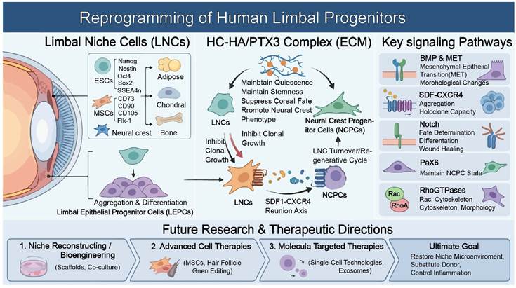
Overview of limbal niche progenitor reprogramming and key regulatory mechanisms. This diagram illustrates the key components and regulatory mechanisms governing limbal niche homeostasis and progenitor reprogramming. (Left) Limbal niche cells (LNCs) and limbal epithelial progenitors (LEPs) reside in the limbal microenvironment. LNCs exhibit multi-lineage differentiation potential (adipogenic, chondrogenic, and osteogenic) and express epithelial stem cell markers (Nanog, Oct4, and Sox2) and stromal progenitor markers (CD73, CD90, CD105, Flk-1), with the capacity to form neural crest-like cells. LNCs interact with LEPs to support their aggregation and differentiation. (Middle) The HC-HA/PTX3 complex (a native extracellular matrix component) maintains LNC quiescence and stemness, suppresses corneal fate commitment, and promotes a neural crest phenotype, thereby supporting the maintenance of niche-derived progenitor cells (NCPCs). Conversely, dysregulated LNCs inhibit clonal growth, whereas the SDF-1/CXCR4 axis facilitates LNC turnover and the regenerative cycle toward NCPCs. (Right) Key signaling pathways regulating limbal progenitors are summarized, including BMP-mediated mesenchymal-epithelial transition (MET) and morphological changes, SDF-1/CXCR4-driven cell aggregation and holoclone formation, Notch signaling for fate determination, differentiation, and wound healing, Pax6 for maintaining the NCPC state, and Rho GTPases (Rac/RhoA) governing cytoskeletal dynamics and morphology. (Bottom) Future research and therapeutic directions include (1) niche reconstruction and bioengineering (scaffolds, co-culture systems), (2) advanced cell therapies (using mesenchymal stem cells, hair follicle cells, and gene editing), and (3) molecularly targeted therapies (single-cell technologies, exosomes), to achieve the ultimate aim of repairing the damaged limbal niche, reducing donor dependency, and controlling inflammation in limbal stem cell deficiency.
Future Directions
Future directions in limbal niche research focus on reconstructing the niche using bioengineered scaffolds, MSCs, alternative cell sources (such as hair follicle- and dental tissue-derived cells), and signaling factors; improving transplantation strategies through advanced single-cell technologies; and exploring novel therapies, including angiogenesis modulation and cellular reprogramming, to prevent corneal disease. Key goals are to restore stem cell function, control inflammation, and develop more effective, less invasive treatments for LSCD.
Key Research Areas:
1. Niche Reconstruction and Bioengineering: a) Mimicking the Niche: To develop artificial ECM or scaffolds that resemble the limbal niche's topography and signals (such as Wnt/BMP) to promote stem cell growth. b) Native Niche Cell Co-Culture: Reconstituting the limbal niche in vitro by co-culturing LESCs with stromal niche cells to understand self-renewal and quiescence.
2. Advanced Cell Therapies: a) MSCs: Using MSCs (from bone marrow, dental pulp, etc.) to modulate inflammation, scarring, and angiogenesis, potentially in co-transplantation with LESCs. b) Alternative Sources: Exploring hair follicles, teeth (dental pulp), and skin as sources of multipotent cells to generate corneal epithelial cells. c) Reprogramming: Reprogramming existing adult corneal cells or pluripotent stem cells into LSC-like cells using gene editing and specific developmental stimuli.
3. Molecular Signaling: a) Single-Cell Technology: Using single-nucleus or single-cell transcriptomics to monitor molecular identity within the human limbal niche and characterize novel markers. b) Signaling Pathways: Investigating how Wnt, BMP, and other signaling pathways regulate LESC self-renewal and fate decisions, as well as the role of persistent inflammation.
4. Therapeutic Factor-Based Approaches: a) Soluble Factors: Delivering specific growth factors as well as cytokines (and extracellular vesicles, such as exosomes) from niche cells to stimulate corneal regeneration. b) Angiogenesis Modulation: Targeting pathological neovascularization, as the limbus normally acts as a barrier to blood vessel invasion.
5. Improving Surgical Techniques: Developing faster, more efficient surgical methods (e.g., simplified mucosal transplantation) and improved scaffolds (such as the amniotic membrane) for cultivated cells.
Overarching Goals: a) Move beyond simple cell transplantation to restore the entire niche microenvironment. b) Reduce reliance on donor corneas and invasive procedures. c) Address inflammation and maintain immune privilege to ensure long-term success.
Conclusion
LNCs are major stromal progenitors in the limbal microenvironment, exhibiting adipogenic, chondrogenic, and osteogenic potential. LNCs express ESC markers, for example, Sox2, Nanog, and Oct4, and mesenchymal progenitor markers, for example, CD90. In addition, LNCs interact with LEPs to support their aggregation and differentiation. Furthermore, the HC-HA/PTX3 complex may maintain LNC quiescence and stemness as well as support niche-derived progenitor cells (NCPCs). Key signaling pathways that maintain limbal progenitor status include BMP-mediated MET, SDF-1/CXCR4-driven cell aggregation and holoclone formation, Notch signaling-mediated fate determination, differentiation, and wound healing, Pax6-mediated maintenance of the NCPC state, and Rho GTPase (Rac/RhoA)-governed cytoskeletal dynamics.
Therefore, understanding the regeneration of adult limbal progenitors and the related mechanisms may pave the way for cell-based therapies of various eye diseases caused by stem cell deficiency or malfunction.
Acknowledgements
Supported by National Natural Science Foundation of China (82471050); High level foreign experts grants of the Ministry of Science and Technology of China (S20240235); Wuhan Optoelectronics National Research Center Open Project (2024WNLOKF002); Open Research Projects of the National Stem Cell Resource Bank for 2026 (NSCRC-26-57).
AI usage statement
No AI usage for preparation, modification, and editing of this manuscript.
Competing Interests
The authors have declared that no competing interest exists.
References
1. Schlotzer-Schrehardt U, Kruse FE. Identification and characterization of limbal stem cells. ExpEye Res. 2005;81:247-64
2. Hayashi R, Yamato M, Sugiyama H, Sumide T, Yang J, Okano T. et al. N-Cadherin is expressed by putative stem/progenitor cells and melanocytes in the human limbal epithelial stem cell niche. Stem Cells. 2007;25:289-96
3. Li S, Zenkel M, Kruse FE, Giessl A, Schlotzer-Schrehardt U. Identification, Isolation, and Characterization of Melanocyte Precursor Cells in the Human Limbal Stroma. Int J Mol Sci. 2022;23:3756
4. Lawrenson JG, Ruskell GL. The structure of corpuscular nerve endings in the limbal conjunctiva of the human eye. J Anat. 1991;177:75-84
5. Chen SY, Hayashida Y, Chen MY, Xie HT, Tseng SC. A new isolation method of human limbal progenitor cells by maintaining close association with their niche cells. Tissue Eng Part C Methods. 2011;17:537-48
6. Su G, Wang W, Xu L, Liu R, Tan Y, Jin B. et al. Isolation and Identification of Limbal Niche Cells. J Vis Exp. 2023 Oct 27:(200). doi: 10.3791/65618
7. Xie HT, Chen SY, Li GG, Tseng SC. Limbal epithelial stem/progenitor cells attract stromal niche cells by SDF-1/CXCR4 signaling to prevent differentiation. Stem Cells. 2011;29:1874-85
8. Espana EM, Romano AC, Kawakita T, Di Pascuale M, Smiddy R, Tseng SC. Novel enzymatic isolation of an entire viable human limbal epithelial sheet. Invest OphthalmolVisSci. 2003;44:4275-81
9. Bhartiya D, Shaikh A, Nagvenkar P, Kasiviswanathan S, Pethe P, Pawani H. et al. Very Small Embryonic-Like Stem Cells with Maximum Regenerative Potential Get Discarded During Cord Blood Banking and Bone Marrow Processing for Autologous Stem Cell Therapy. Stem Cells Dev. 2011;21:1-6
10. Li GG, Chen SY, Xie HT, Zhu YT, Tseng SC. Angiogenesis potential of human limbal stromal niche cells. Investigative ophthalmology & visual science. 2012;53:3357-67
11. Nikolova G, Strilic B, Lammert E. The vascular niche and its basement membrane. Trends Cell Biol. 2007;17:19-25
12. Kopp HG, Avecilla ST, Hooper AT, Rafii S. The bone marrow vascular niche: home of HSC differentiation and mobilization. Physiology(Bethesda). 2005;20:349-56
13. Wang W, Li S, Xu L, Jiang M, Li X, Zhang Y. et al. Differential Gene Expression between Limbal Niche Progenitors and Bone Marrow Derived Mesenchymal Stem Cells. Int J Med Sci. 2020;17:549-57
14. Fujiwara H, Hayashi Y, Sanzen N, Kobayashi R, Weber CN, Emoto T. et al. Regulation of mesodermal differentiation of mouse embryonic stem cells by basement membranes. JBiolChem. 2007;282:29701-11
15. Zhu H, Wang W, Tan Y, Su G, Xu L, Jiang ML. et al. Limbal Niche Cells and Three-Dimensional Matrigel-Induced Dedifferentiation of Mature Corneal Epithelial Cells. Investigative ophthalmology & visual science. 2022;63:1
16. Lindner U, Kramer J, Behrends J, Driller B, Wendler NO, Boehrnsen F. et al. Improved proliferation and differentiation capacity of human mesenchymal stromal cells cultured with basement-membrane extracellular matrix proteins. Cytotherapy. 2010;12:992-1005
17. Matsubara T, Tsutsumi S, Pan H, Hiraoka H, Oda R, Nishimura M. et al. A new technique to expand human mesenchymal stem cells using basement membrane extracellular matrix. BiochemBiophysResCommun. 2004;313:503-8
18. Garrett RW, Emerson SG. Bone and blood vessels: the hard and the soft of hematopoietic stem cell niches. Cell Stem Cell. 2009;4:503-6
19. Shen Q, Goderie SK, Jin L, Karanth N, Sun Y, Abramova N. et al. Endothelial cells stimulate self-renewal and expand neurogenesis of neural stem cells. Science. 2004;304:1338-40
20. Tavazoie M, van d V, Silva-Vargas V, Louissaint M, Colonna L, Zaidi B. et al. A specialized vascular niche for adult neural stem cells. Cell Stem Cell. 2008;3:279-88
21. Christov C, Chretien F, bou-Khalil R, Bassez G, Vallet G, Authier FJ. et al. Muscle satellite cells and endothelial cells: close neighbors and privileged partners. MolBiolCell. 2007;18:1397-409
22. Lammert E, Cleaver O, Melton D. Induction of pancreatic differentiation by signals from blood vessels. Science. 2001;294:564-7
23. Whitman MC, Fan W, Rela L, Rodriguez-Gil DJ, Greer CA. Blood vessels form a migratory scaffold in the rostral migratory stream. JComp Neurol. 2009;516:94-104
24. Kolli S, Bojic S, Ghareeb AE, Kurzawa-Akanbi M, Figueiredo FC, Lako M. The Role of Nerve Growth Factor in Maintaining Proliferative Capacity, Colony-Forming Efficiency, and the Limbal Stem Cell Phenotype. Stem Cells. 2019;37:139-49
25. Dore-Duffy P, Katychev A, Wang X, Van BE. CNS microvascular pericytes exhibit multipotential stem cell activity. JCerebBlood Flow Metab. 2006;26:613-24
26. Shi S, Gronthos S. Perivascular niche of postnatal mesenchymal stem cells in human bone marrow and dental pulp. JBone MinerRes. 2003;18:696-704
27. Crisan M, Yap S, Casteilla L, Chen CW, Corselli M, Park TS. et al. A perivascular origin for mesenchymal stem cells in multiple human organs. Cell Stem Cell. 2008;3:301-13
28. Crisan M, Deasy B, Gavina M, Zheng B, Huard J, Lazzari L. et al. Purification and long-term culture of multipotent progenitor cells affiliated with the walls of human blood vessels: myoendothelial cells and pericytes. Methods Cell Biol. 2008;86:295-309
29. da Silva ML, Chagastelles PC, Nardi NB. Mesenchymal stem cells reside in virtually all post-natal organs and tissues. JCell Sci. 2006;119:2204-13
30. Schraufstatter IU, Discipio RG, Khaldoyanidi S. Mesenchymal stem cells and their microenvironment. Front Biosci. 2011;17:2271-88
31. Prina E, Amer MH, Sidney L, Tromayer M, Moore J, Liska R. et al. Bioinspired Precision Engineering of Three-Dimensional Epithelial Stem Cell Microniches. Adv Biosyst. 2020;4:e2000016
32. Behaegel J, Zakaria N, Tassignon MJ, Leysen I, Bock F, Koppen C. et al. Short- and Long-Term Results of Xenogeneic-Free Cultivated Autologous and Allogeneic Limbal Epithelial Stem Cell Transplantations. Cornea. 2019;38:1543-9
33. Zhu L, Zhang W, Zhu J, Chen C, Mo K, Guo H. et al. Cotransplantation of Limbal Epithelial and Stromal Cells for Ocular Surface Reconstruction. Ophthalmol Sci. 2022;2:100148
34. Yazdanpanah G, Haq Z, Kang K, Jabbehdari S, Rosenblatt ML, Djalilian AR. Strategies for reconstructing the limbal stem cell niche. The ocular surface. 2019;17:230-40
35. Polisetti N, Roschinski B, Schlotzer-Schrehardt U, Maier P, Schlunck G, Reinhard T. A Decellularized Human Limbal Scaffold for Limbal Stem Cell Niche Reconstruction. Int J Mol Sci. 2021;22:10067
36. Singh V, Tiwari A, Kethiri AR, Sangwan VS. Current perspectives of limbal-derived stem cells and its application in ocular surface regeneration and limbal stem cell transplantation. Stem Cells Transl Med. 2021;10:1121-8
37. Tan Y, Wang W, Xu L, Zhou X, Shen J, Zhou T. et al. Decellularized human amniotic member hydrogel promotes limbal stem cells proliferation. Colloids and surfaces B, Biointerfaces. 2025;252:114656
38. Tseng SCG. Amniotic membrane transplantation for ocular surface reconstruction. Bioscience Rep. 2002;21:481-9
39. Dua HS, Gomes JA, King AJ, Maharajan VS. The amniotic membrane in ophthalmology. SurvOphthalmol. 2004;49:51-77
40. Bouchard CS, John T. Amniotic membrane transplantation in the management of severe ocular surface disease: indications and outcomes. OculSurf. 2004;2:201-11
41. Tseng TCG. Niche Regulation of Limbal Epithelial Stem Cells: HC-HA/PTX3 as Surrogate Matrix Niche. Experimental eye research. 2020;199:108181
42. Robertson SYT, Roberts JS, Deng SX. Regulation of Limbal Epithelial Stem Cells: Importance of the Niche. Int J Mol Sci. 2021;22:11975
43. Moreno IY, Parsaie A, Gesteira TF, Coulson-Thomas VJ. Characterization of the Limbal Epithelial Stem Cell Niche. Investigative ophthalmology & visual science. 2023;64:48
44. Bonnet C, Gonzalez S, Roberts JS, Robertson SYT, Ruiz M, Zheng J. et al. Human limbal epithelial stem cell regulation, bioengineering and function. Prog Retin Eye Res. 2021;85:100956
45. Heiligenhaus A, Bauer D, Meller D, Steuhl KP, Tseng SC. Improvement of HSV-1 necrotizing keratitis with amniotic membrane transplantation. Investigative ophthalmology & visual science. 2001;42:1969-74
46. Heiligenhaus A, Li H, Hernandez Galindo EE, Koch JM, Steuhl KP, Meller D. Management of acute ulcerative and necrotising herpes simplex and zoster keratitis with amniotic membrane transplantation. BrJ Ophthalmol. 2003;87:1215-9
47. Bauer D, Wasmuth S, Hennig M, Baehler H, Steuhl KP, Heiligenhaus A. Amniotic membrane transplantation induces apoptosis in T lymphocytes in murine corneas with experimental herpetic stromal keratitis. Investigative ophthalmology & visual science. 2009;50:3188-98
48. He H, Li W, Tseng DY, Zhang S, Chen SY, Day AJ. et al. Biochemical characterization and function of complexes formed by hyaluronan and the heavy chains of inter-alpha-inhibitor (HC*HA) purified from extracts of human amniotic membrane. J Biol Chem. 2009;284:20136-46
49. He H, Li W, Chen SY, Zhang S, Chen YT, Hayashida Y. et al. Suppression of activation and induction of apoptosis in RAW264.7 cells by amniotic membrane extract. Investigative ophthalmology & visual science. 2008;49:4468-75
50. Shay E, He H, Sakurai S, Tseng SC. Inhibition of angiogenesis by HC.HA, a complex of hyaluronan and the heavy chain of inter-alpha-inhibitor, purified from human amniotic membrane. Invest OphthalmolVisSci. 2011;52:2669-78
51. Garlanda C, Bottazzi B, Bastone A, Mantovani A. Pentraxins at the crossroads between innate immunity, inflammation, matrix deposition, and female fertility. AnnuRevImmunol. 2005;23:337-66
52. Colon E, Shytuhina A, Cowman MK, Band PA, Sanggaard KW, Enghild JJ. et al. Transfer of inter-alpha-inhibitor heavy chains to hyaluronan by surface-linked hyaluronan-TSG-6 complexes. JBiolChem. 2009;284:2320-31
53. Lee TH, Klampfer L, Shows TB, Vilcek J. Transcriptional regulation of TSG6, a tumor necrosis factor- and interleukin-1-inducible primary response gene coding for a secreted hyaluronan-binding protein. JBiolChem. 1993;268:6154-60
54. Wisniewski HG, Vilcek J. TSG-6: an IL-1/TNF-inducible protein with anti-inflammatory activity. Cytokine Growth Factor Rev. 1997;8:143-56
55. Lee TH, Wisniewski H-G, Vilcek J. A novel secretory tumor necrosis factor-inducible protein (TSG-6) is a member of the family of hyaluronate binding protein, closely related to the adhesion receptor CD44. JCell Biol. 1992;116:545-57
56. Lee TH, Lee GW, Ziff EB, Vilcek J. Isolation and characterization of eight tumor necrosis factor-induced gene sequences from human fibroblasts. MolCell Biol. 1990;10:1982-8
57. Xie HT, Chen SY, Li GG, Tseng SC. Isolation and expansion of human limbal stromal niche cells. Investigative ophthalmology & visual science. 2012;53:279-86
58. Li GG, Zhu YT, Xie HT, Chen SY, Tseng SC. Mesenchymal stem cells derived from human limbal niche cells. Investigative ophthalmology & visual science. 2012;53:5686-97
59. Han B, Chen SY, Zhu YT, Tseng SC. Integration of BMP/Wnt signaling to control clonal growth of limbal epithelial progenitor cells by niche cells. Stem Cell Res. 2014;12:562-73
60. Chen SY, Han B, Zhu YT, Mahabole M, Huang J, Beebe DC. et al. HC-HA/PTX3 Purified from Amniotic Membrane Promotes BMP Signaling in Limbal Niche Cells to Maintain Quiescence of Limbal Epithelial Progenitor/Stem Cells. Stem Cells. 2015;33:3341-55
61. Sangwan VS, Basu S, MacNeil S, Balasubramanian D. Simple limbal epithelial transplantation (SLET): a novel surgical technique for the treatment of unilateral limbal stem cell deficiency. Br J Ophthalmol. 2012;96:931-4
62. Amescua G, Atallah M, Nikpoor N, Galor A, Perez V. Modified simple limbal epithelial transplantation using cryopreserved amniotic membrane for unilateral limbal stem cell deficiency. Am J Ophthalmol. 2014;158:469-75
63. Tseng SC, Chen SY, Shen YC, Chen WL, Hu FR. Critical appraisal of ex vivo expansion of human limbal epithelial stem cells. Curr Mol Med. 2010;10:841-50
64. Galindo S, Lopez-Paniagua M, Mata A, Herreras JM, Garcia-Vazquez C, Marcenido B. et al. Bone marrow-versus adipose tissue-derived mesenchymal stem cells for corneal failure in an experimental model of limbal stem cell deficiency. Experimental eye research. 2026;262:110737
65. Streit T, Ullrich K, Geerling G, Spaniol K. [Limbal diseases]. Ophthalmologie. 2026;123:75-89
66. Park M, Pandzic E, Males J, Di Girolamo N. Mechanistic insights into conjunctivalization in limbal stem cell deficiency using a lineage tracing approach. The ocular surface. 2026;39:21-33
67. Takahashi K, Yamanaka S. Induction of pluripotent stem cells from mouse embryonic and adult fibroblast cultures by defined factors. Cell. 2006;126:663-76
68. Chen T, Yuan D, Wei B, Jiang J, Kang J, Ling K. et al. E-cadherin-mediated cell-cell contact is critical for induced pluripotent stem cell generation. Stem Cells. 2010;28:1315-25
69. Li L, Wang BH, Wang S, Moalim-Nour L, Mohib K, Lohnes D. et al. Individual cell movement, asymmetric colony expansion, rho-associated kinase, and E-cadherin impact the clonogenicity of human embryonic stem cells. Biophys J. 2010;98:2442-51
70. Samavarchi-Tehrani P, Golipour A, David L, Sung HK, Beyer TA, Datti A. et al. Functional genomics reveals a BMP-driven mesenchymal-to-epithelial transition in the initiation of somatic cell reprogramming. Cell Stem Cell. 2010;7:64-77
71. Li R, Liang J, Ni S, Zhou T, Qing X, Li H. et al. A mesenchymal-to-epithelial transition initiates and is required for the nuclear reprogramming of mouse fibroblasts. Cell Stem Cell. 2010;7:51-63
72. Larue L, Ohsugi M, Hirchenhain J, Kemler R. E-cadherin null mutant embryos fail to form a trophectoderm epithelium. ProcNatlAcadSciUSA. 1994;91:8263-7
73. Das S, Becker BN, Hoffmann FM, Mertz JE. Complete reversal of epithelial to mesenchymal transition requires inhibition of both ZEB expression and the Rho pathway. BMCCell Biol. 2009;10:94
74. Zhu YT, Chen HC, Chen SY, Tseng SC. Nuclear p120 catenin unlocks mitotic block of contact-inhibited human corneal endothelial monolayers without disrupting adherent junctions. JCell Sci. 2012;125:3636-48
75. Zhu YT, Li F, Han B, Tighe S, Zhang S, Chen SY. et al. Activation of RhoA-ROCK-BMP signaling reprograms adult human corneal endothelial cells. JCell Biol. 2014;206:799-811
76. Chen S, Zhu Q, Sun H, Zhang Y, Tighe S, Xu L. et al. Advances in culture, expansion and mechanistic studies of corneal endothelial cells: a systematic review. J Biomed Sci. 2019;26:2
77. Sun H, Zhu Q, Guo P, Zhang Y, Tighe S, Zhu Y. Trabecular meshwork cells are a valuable resource for cellular therapy of glaucoma. J Cell Mol Med. 2019;23:1678-86
78. Yang X, Xu Z, Li D, Cheng S, Fan K, Li C. et al. Cell surface nucleolin is crucial in the activation of the CXCL12/CXCR4 signaling pathway. Tumour biology: the journal of the International Society for Oncodevelopmental Biology and Medicine. 2014;35:333-8
79. David L, Polo JM. Phases of reprogramming. Stem Cell Res. 2014;12:754-61
80. Hayashi Y, Hsiao EC, Sami S, Lancero M, Schlieve CR, Nguyen T. et al. BMP-SMAD-ID promotes reprogramming to pluripotency by inhibiting p16/INK4A-dependent senescence. Proc Natl Acad Sci U S A. 2016;113:13057-62
81. Mikkelsen TS, Hanna J, Zhang X, Ku M, Wernig M, Schorderet P. et al. Dissecting direct reprogramming through integrative genomic analysis. Nature. 2008;454:49-55
82. Li M, Yu J, Li Y, Li D, Yan D, Ruan Q. CXCR4+ progenitors derived from bone mesenchymal stem cells differentiate into endothelial cells capable of vascular repair after arterial injury. Cell Reprogram. 2010;12:405-15
83. Hong H, Takahashi K, Ichisaka T, Aoi T, Kanagawa O, Nakagawa M. et al. Suppression of induced pluripotent stem cell generation by the p53-p21 pathway. Nature. 2009;460:1132-5
84. Marion RM, Strati K, Li H, Murga M, Blanco R, Ortega S. et al. A p53-mediated DNA damage response limits reprogramming to ensure iPS cell genomic integrity. Nature. 2009;460:1149-53
85. Utikal J, Polo JM, Stadtfeld M, Maherali N, Kulalert W, Walsh RM. et al. Immortalization eliminates a roadblock during cellular reprogramming into iPS cells. Nature. 2009;460:1145-8
86. Jin B, Su G, Zhou X, Xu L, Wang W, Zhou T. et al. Basic Fibroblast Growth Factor Supports the Function of Limbal Niche Cells via the Wnt/beta-Catenin Pathway. Journal of ocular pharmacology and therapeutics: the official journal of the Association for Ocular Pharmacology and Therapeutics. 2024;40:571-80
87. Spradling A, Drummond-Barbosa D, Kai T. Stem cells find their niche. Nature. 2001;414:98-104
88. Fuchs E, Tumbar T, Guasch G. Socializing with the neighbors: stem cells and their niche. Cell. 2004;116:769-78
89. Moore KA, Lemischka IR. Stem cells and their niches. Science. 2006;311:1880-5
90. Zhang J, He XC, Tong WG, Johnson T, Wiedemann LM, Mishina Y. et al. Bone morphogenetic protein signaling inhibits hair follicle anagen induction by restricting epithelial stem/progenitor cell activation and expansion. Stem Cells. 2006;24:2826-39
91. Kobielak K, Stokes N, de la CJ, Polak L, Fuchs E. Loss of a quiescent niche but not follicle stem cells in the absence of bone morphogenetic protein signaling. ProcNatlAcadSciUSA. 2007;104:10063-8
92. Jamora C, DasGupta R, Kocieniewski P, Fuchs E. Links between signal transduction, transcription and adhesion in epithelial bud development. Nature. 2003;422:317-22
93. Gregorieff A, Pinto D, Begthel H, Destree O, Kielman M, Clevers H. Expression pattern of Wnt signaling components in the adult intestine. Gastroenterology. 2005;129:626-38
94. van Es JH, Jay P, Gregorieff A, van Gijn ME, Jonkheer S, Hatzis P. et al. Wnt signalling induces maturation of Paneth cells in intestinal crypts. NatCell Biol. 2005;7:381-6
95. Gregory KE, Ono RN, Charbonneau NL, Kuo CL, Keene DR, Bachinger HP. et al. The prodomain of BMP-7 targets the BMP-7 complex to the extracellular matrix. JBiolChem. 2005;280:27970-80
96. Pereira RC, Economides AN, Canalis E. Bone morphogenetic proteins induce gremlin, a protein that limits their activity in osteoblasts. Endocrinology. 2000;141:4558-63
97. Khokha MK, Hsu D, Brunet LJ, Dionne MS, Harland RM. Gremlin is the BMP antagonist required for maintenance of Shh and Fgf signals during limb patterning. NatGenet. 2003;34:303-7
98. Gazzerro E, Gangji V, Canalis E. Bone morphogenetic proteins induce the expression of noggin, which limits their activity in cultured rat osteoblasts. JClinInvest. 1998;102:2106-14
99. Brunet LJ, McMahon JA, McMahon AP, Harland RM. Noggin, cartilage morphogenesis, and joint formation in the mammalian skeleton. Science. 1998;280:1455-7
100. Xu RH, Peck RM, Li DS, Feng X, Ludwig T, Thomson JA. Basic FGF and suppression of BMP signaling sustain undifferentiated proliferation of human ES cells. NatMethods. 2005;2:185-90
101. Rosen V. BMP and BMP inhibitors in bone. AnnNYAcadSci. 2006;1068:19-25
102. Bouwmeester T, Kim S, Sasai Y, Lu B, De Robertis EM. Cerberus is a head-inducing secreted factor expressed in the anterior endoderm of Spemann's organizer. Nature. 1996;382:595-601
103. Bachiller D, Klingensmith J, Kemp C, Belo JA, Anderson RM, May SR. et al. The organizer factors Chordin and Noggin are required for mouse forebrain development. Nature. 2000;403:658-61
104. Simmons DG, Kennedy TG. Uterine sensitization-associated gene-1: a novel gene induced within the rat endometrium at the time of uterine receptivity/sensitization for the decidual cell reaction. BiolReprod. 2002;67:1638-45
105. Marques S, Borges AC, Silva AC, Freitas S, Cordenonsi M, Belo JA. The activity of the Nodal antagonist Cerl-2 in the mouse node is required for correct L/R body axis. Genes Dev. 2004;18:2342-7
106. Walsh DW, Godson C, Brazil DP, Martin F. Extracellular BMP-antagonist regulation in development and disease: tied up in knots. Trends Cell Biol. 2010;20:244-56
107. Wiater E, Vale W. Inhibin is an antagonist of bone morphogenetic protein signaling. JBiolChem. 2003;278:7934-41
108. Gamer LW, Nove J, Levin M, Rosen V. BMP-3 is a novel inhibitor of both activin and BMP-4 signaling in Xenopus embryos. DevBiol. 2005;285:156-68
109. Salbreux G, Charras G, Paluch E. Actin cortex mechanics and cellular morphogenesis. Trends Cell Biol. 2012;22:536-45
110. He S, Liu X, Yang Y, Huang W, Xu S, Yang S. et al. Mechanisms of transforming growth factor beta(1)/Smad signalling mediated by mitogen-activated protein kinase pathways in keloid fibroblasts. Br J Dermatol. 2010;162:538-46
111. Rauzi M, Lenne PF, Lecuit T. Planar polarized actomyosin contractile flows control epithelial junction remodelling. Nature. 2010;468:1110-4
112. Levayer R, Pelissier-Monier A, Lecuit T. Spatial regulation of Dia and Myosin-II by RhoGEF2 controls initiation of E-cadherin endocytosis during epithelial morphogenesis. Nat Cell Biol. 2011;13:529-40
113. Mason FM, Tworoger M, Martin AC. Apical domain polarization localizes actin-myosin activity to drive ratchet-like apical constriction. Nat Cell Biol. 2013;15:926-36
114. Kasza KE, Farrell DL, Zallen JA. Spatiotemporal control of epithelial remodeling by regulated myosin phosphorylation. Proc Natl Acad Sci U S A. 2014;111:11732-7
115. Vasquez CG, Tworoger M, Martin AC. Dynamic myosin phosphorylation regulates contractile pulses and tissue integrity during epithelial morphogenesis. J Cell Biol. 2014;206:435-50
116. Munjal A, Philippe JM, Munro E, Lecuit T. A self-organized biomechanical network drives shape changes during tissue morphogenesis. Nature. 2015;524:351-5
117. Jaffe AB, Hall A, Schmidt A. Association of CNK1 with Rho guanine nucleotide exchange factors controls signaling specificity downstream of Rho. Curr Biol. 2005;15:405-12
118. Barrett K, Leptin M, Settleman J. The Rho GTPase and a putative RhoGEF mediate a signaling pathway for the cell shape changes in Drosophila gastrulation. Cell. 1997;91:905-15
119. Smallhorn M, Murray MJ, Saint R. The epithelial-mesenchymal transition of the Drosophila mesoderm requires the Rho GTP exchange factor Pebble. Development. 2004;131:2641-51
120. Fox DT, Peifer M. Abelson kinase (Abl) and RhoGEF2 regulate actin organization during cell constriction in Drosophila. Development. 2007;134:567-78
121. Sawyer JM, Harrell JR, Shemer G, Sullivan-Brown J, Roh-Johnson M, Goldstein B. Apical constriction: a cell shape change that can drive morphogenesis. Dev Biol. 2010;341:5-19
122. Martin AC, Goldstein B. Apical constriction: themes and variations on a cellular mechanism driving morphogenesis. Development. 2014;141:1987-98
123. Kolsch V, Seher T, Fernandez-Ballester GJ, Serrano L, Leptin M. Control of Drosophila gastrulation by apical localization of adherens junctions and RhoGEF2. Science. 2007;315:384-6
124. Manning AJ, Peters KA, Peifer M, Rogers SL. Regulation of epithelial morphogenesis by the G protein-coupled receptor mist and its ligand fog. Sci Signal. 2013;6:ra98
125. Kerridge S, Munjal A, Philippe JM, Jha A, de las Bayonas AG, Saurin AJ. et al. Modular activation of Rho1 by GPCR signalling imparts polarized myosin II activation during morphogenesis. Nat Cell Biol. 2016;18:261-70
126. Martin AC, Kaschube M, Wieschaus EF. Pulsed contractions of an actin-myosin network drive apical constriction. Nature. 2009;457:495-9
127. Munro P, Flatau G, Doye A, Boyer L, Oregioni O, Mege JL. et al. Activation and proteasomal degradation of rho GTPases by cytotoxic necrotizing factor-1 elicit a controlled inflammatory response. J Biol Chem. 2004;279:35849-57
128. Mason FM, Xie S, Vasquez CG, Tworoger M, Martin AC. RhoA GTPase inhibition organizes contraction during epithelial morphogenesis. J Cell Biol. 2016;214:603-17
129. Teo JL, Yap AS. Cycling Rho for tissue contraction. J Cell Biol. 2016;214:495-8
130. Parri M, Chiarugi P. Rac and Rho GTPases in cancer cell motility control. Cell Commun Signal. 2010;8:23
131. Askari AT, Unzek S, Popovic ZB, Goldman CK, Forudi F, Kiedrowski M. et al. Effect of stromal-cell-derived factor 1 on stem-cell homing and tissue regeneration in ischaemic cardiomyopathy. Lancet (London, England). 2003;362:697-703
132. Rosenkranz K, Kumbruch S, Lebermann K, Marschner K, Jensen A, Dermietzel R. et al. The chemokine SDF-1/CXCL12 contributes to the 'homing' of umbilical cord blood cells to a hypoxic-ischemic lesion in the rat brain. J Neurosci Res. 2010;88:1223-33
133. Stokman G, Stroo I, Claessen N, Teske GJ, Florquin S, Leemans JC. SDF-1 provides morphological and functional protection against renal ischaemia/reperfusion injury. Nephrol Dial Transplant. 2010;25:3852-9
134. Deglurkar I, Mal N, Mills WR, Popovic ZB, McCarthy P, Blackstone EH. et al. Mechanical and electrical effects of cell-based gene therapy for ischemic cardiomyopathy are independent. Hum Gene Ther. 2006;17:1144-51
135. Unzek S, Zhang M, Mal N, Mills WR, Laurita KR, Penn MS. SDF-1 recruits cardiac stem cell-like cells that depolarize in vivo. Cell Transplant. 2007;16:879-86
136. Zhang XQ, Yamada S, Barry WH. Ranolazine inhibits an oxidative stress-induced increase in myocyte sodium and calcium loading during simulated-demand ischemia. J Cardiovasc Pharmacol. 2008;51:443-9
137. Chen SY, Zhu Y, Zhang Y, Hsu D, Tseng SCG. HC-HA/PTX3 from amniotic membrane reverts senescent limbal niche cells to Pax6+ neural crest progenitors to support limbal epithelial progenitors. Stem Cells. 2021;39:280-95
138. Tseng SCG, Chen SY, Mead OG, Tighe S. Niche regulation of limbal epithelial stem cells: HC-HA/PTX3 as surrogate matrix niche. Experimental eye research. 2020;199:108181
139. Zhu YT, Li F, Zhang Y, Chen SY, Tighe S, Lin SY. et al. HC-HA/PTX3 Purified From Human Amniotic Membrane Reverts Human Corneal Fibroblasts and Myofibroblasts to Keratocytes by Activating BMP Signaling. Investigative ophthalmology & visual science. 2020;61:62
140. Aghazadeh S, Peng Q, Dardmeh F, Ostergaard Hjortdal J, Zachar V, Alipour H. The impact of the limbal niche interactions on the self-renewal capability of limbal epithelial stem cells. Front Cell Dev Biol. 2025;13:1667309
141. Charo IF, Ransohoff RM. The many roles of chemokines and chemokine receptors in inflammation. NEnglJMed. 2006;354:610-21
142. Burns JM, Summers BC, Wang Y, Melikian A, Berahovich R, Miao Z. et al. A novel chemokine receptor for SDF-1 and I-TAC involved in cell survival, cell adhesion, and tumor development. JExpMed. 2006;203:2201-13
143. Oberlin E, Amara A, Bachelerie F, Bessia C, Virelizier JL, renzana-Seisdedos F. et al. The CXC chemokine SDF-1 is the ligand for LESTR/fusin and prevents infection by T-cell-line-adapted HIV-1. Nature. 1996;382:833-5
144. Sengupta N, Afzal A, Caballero S, Chang KH, Shaw LC, Pang JJ. et al. Paracrine modulation of CXCR4 by IGF-1 and VEGF: implications for choroidal neovascularization. Invest OphthalmolVisSci. 2010;51:2697-704
145. Liu H, Liu S, Li Y, Wang X, Xue W, Ge G. et al. The role of SDF-1-CXCR4/CXCR7 axis in the therapeutic effects of hypoxia-preconditioned mesenchymal stem cells for renal ischemia/reperfusion injury. PLoSOne. 2012;7:e34608
146. Nakayama T, Mutsuga N, Tosato G. FGF2 posttranscriptionally down-regulates expression of SDF1 in bone marrow stromal cells through FGFR1 IIIc. Blood. 2007;109:1363-72
147. Florin L, Maas-Szabowski N, Werner S, Szabowski A, Angel P. Increased keratinocyte proliferation by JUN-dependent expression of PTN and SDF-1 in fibroblasts. JCell Sci. 2005;118:1981-9
148. Vauclair S, Majo F, Durham AD, Ghyselinck NB, Barrandon Y, Radtke F. Corneal epithelial cell fate is maintained during repair by Notch1 signaling via the regulation of vitamin A metabolism. DevCell. 2007;13:242-53
149. Ma A, Boulton M, Zhao B, Connon C, Cai J, Albon J. A role for notch signaling in human corneal epithelial cell differentiation and proliferation. Invest OphthalmolVisSci. 2007;48:3576-85
150. Lu H, Lu Q, Zheng Y, Li Q. Notch signaling promotes the corneal epithelium wound healing. Mol Vis. 2012;18:403-11
151. Altshuler A, Amitai-Lange A, Tarazi N, Dey S, Strinkovsky L, Hadad-Porat S. et al. Discrete limbal epithelial stem cell populations mediate corneal homeostasis and wound healing. Cell Stem Cell. 2021;28:1248-61
152. Nakamura T, Ohtsuka T, Sekiyama E, Cooper LJ, Kokubu H, Fullwood NJ. et al. Hes1 regulates corneal development and the function of corneal epithelial stem/progenitor cells. Stem Cells. 2008;26:1265-74
153. Ma DH, Lai JY, Cheng HY, Tsai CC, Yeh LK. Carbodiimide cross-linked amniotic membranes for cultivation of limbal epithelial cells. Biomaterials. 2010;31:6647-58
154. Djalilian AR, Namavari A, Ito A, Balali S, Afshar A, Lavker RM. et al. Down-regulation of Notch signaling during corneal epithelial proliferation. MolVis. 2008;14:1041-9
155. Li J, Chen SY, Zhao XY, Zhang MC, Xie HT. Rat Limbal Niche Cells Prevent Epithelial Stem/Progenitor Cells From Differentiation and Proliferation by Inhibiting Notch Signaling Pathway In Vitro. Investigative ophthalmology & visual science. 2017;58:2968-76
156. Sanalkumar R, Dhanesh SB, James J. Non-canonical activation of Notch signaling/target genes in vertebrates. Cell Mol Life Sci. 2010;67:2957-68
157. Salama-Cohen P, Arevalo MA, Meier J, Grantyn R, Rodriguez-Tebar A. NGF controls dendrite development in hippocampal neurons by binding to p75NTR and modulating the cellular targets of Notch. Mol Biol Cell. 2005;16:339-47
158. Hashimoto T, Zhang XM, Chen BY, Yang XJ. VEGF activates divergent intracellular signaling components to regulate retinal progenitor cell proliferation and neuronal differentiation. Development. 2006;133:2201-10
159. Ichi S, Boshnjaku V, Shen YW, Mania-Farnell B, Ahlgren S, Sapru S. et al. Role of Pax3 acetylation in the regulation of Hes1 and Neurog2. Mol Biol Cell. 2011;22:503-12
160. Ohtsuka T, Sakamoto M, Guillemot F, Kageyama R. Roles of the basic helix-loop-helix genes Hes1 and Hes5 in expansion of neural stem cells of the developing brain. J Biol Chem. 2001;276:30467-74
161. Baek JH, Hatakeyama J, Sakamoto S, Ohtsuka T, Kageyama R. Persistent and high levels of Hes1 expression regulate boundary formation in the developing central nervous system. Development. 2006;133:2467-76
162. Hirata H, Yoshiura S, Ohtsuka T, Bessho Y, Harada T, Yoshikawa K. et al. Oscillatory expression of the bHLH factor Hes1 regulated by a negative feedback loop. Science. 2002;298:840-3
163. Shimojo H, Ohtsuka T, Kageyama R. Oscillations in notch signaling regulate maintenance of neural progenitors. Neuron. 2008;58:52-64
164. Borggrefe T, Oswald F. The Notch signaling pathway: transcriptional regulation at Notch target genes. Cell Mol Life Sci. 2009;66:1631-46
165. Lee JH, Suk J, Park J, Kim SB, Kwak SS, Kim JW. et al. Notch signal activates hypoxia pathway through HES1-dependent SRC/signal transducers and activators of transcription 3 pathway. Molecular cancer research: MCR. 2009;7:1663-71
166. Polisetti N, Schlunck G, Reinhard T. PAX6 Expression Patterns in the Adult Human Limbal Stem Cell Niche. Cells. 2023;12:400
167. Chen SY, Cheng AMS, Zhang Y, Zhu YT, He H, Mahabole M. et al. Pax 6 Controls Neural Crest Potential of Limbal Niche Cells to Support Self-Renewal of Limbal Epithelial Stem Cells. Sci Rep. 2019;9:9763
168. Ghareeb AE, Lako M, Figueiredo FC. Recent Advances in Stem Cell Therapy for Limbal Stem Cell Deficiency: A Narrative Review. Ophthalmol Ther. 2020;9:809-31
169. Chiriac A, Nelson TJ, Faustino RS, Behfar A, Terzic A. Cardiogenic induction of pluripotent stem cells streamlined through a conserved SDF-1/VEGF/BMP2 integrated network. PLoSOne. 2010;5:e9943
170. Ping YF, Yao XH, Jiang JY, Zhao LT, Yu SC, Jiang T. et al. The chemokine CXCL12 and its receptor CXCR4 promote glioma stem cell-mediated VEGF production and tumour angiogenesis via PI3K/AKT signalling. JPathol. 2011;224:344-54
171. El Hadri K, Mahmood DF, Couchie D, Jguirim-Souissi I, Genze F, Diderot V. et al. Thioredoxin-1 promotes anti-inflammatory macrophages of the M2 phenotype and antagonizes atherosclerosis. Arterioscler Thromb Vasc Biol. 2012;32:1445-52
172. Toone WM, Morgan BA, Jones N. Redox control of AP-1-like factors in yeast and beyond. Oncogene. 2001;20:2336-46
173. Nishiyama A, Matsui M, Iwata S, Hirota K, Masutani H, Nakamura H. et al. Identification of thioredoxin-binding protein-2/vitamin D(3) up-regulated protein 1 as a negative regulator of thioredoxin function and expression. J Biol Chem. 1999;274:21645-50
174. Lee J, Ku T, Yu H, Chong K, Ryu SW, Choi K. et al. Blockade of VEGF-A suppresses tumor growth via inhibition of autocrine signaling through FAK and AKT. Cancer letters. 2012;318:221-5
175. Vogelmann R, Nguyen-Tat MD, Giehl K, Adler G, Wedlich D, Menke A. TGFbeta-induced downregulation of E-cadherin-based cell-cell adhesion depends on PI3-kinase and PTEN. J Cell Sci. 2005;118:4901-12
176. Mundy GR. The effects of TGF-beta on bone. Ciba Found Symp. 1991;157:137-43 discussion 43-51
177. Rosenfeld CS, Bolwell B, LeFever A, Taylor R, List A, Fay J. et al. Comparison of four cytokine regimens for mobilization of peripheral blood stem cells: IL-3 alone and combined with GM-CSF or G-CSF. Bone Marrow Transplant. 1996;17:179-83
178. Schwarzenberger P, Huang W, Oliver P, Byrne P, La Russa V, Zhang Z. et al. Il-17 mobilizes peripheral blood stem cells with short- and long-term repopulating ability in mice. J Immunol. 2001;167:2081-6
179. Craddock CF, Nakamoto B, Andrews RG, Priestley GV, Papayannopoulou T. Antibodies to VLA4 integrin mobilize long-term repopulating cells and augment cytokine-induced mobilization in primates and mice. Blood. 1997;90:4779-88
180. Walenta KL, Bettink S, Bohm M, Friedrich EB. Differential chemokine receptor expression regulates functional specialization of endothelial progenitor cell subpopulations. Basic Res Cardiol. 2011;106:299-305
181. Peled A, Petit I, Kollet O, Magid M, Ponomaryov T, Byk T. et al. Dependence of human stem cell engraftment and repopulation of NOD/SCID mice on CXCR4. Science. 1999;283:845-8
182. Calderon L, Boehm T. Synergistic, context-dependent, and hierarchical functions of epithelial components in thymic microenvironments. Cell. 2012;149:159-72
183. Bhattacharya S, Mukherjee A, Pisano S, Dimri S, Knaane E, Altshuler A. et al. The biophysical property of the limbal niche maintains stemness through YAP. Cell Death Differ. 2023;30:1601-14
184. Gouveia RM, Lepert G, Gupta S, Mohan RR, Paterson C, Connon CJ. Assessment of corneal substrate biomechanics and its effect on epithelial stem cell maintenance and differentiation. Nat Commun. 2019;10:1496
185. Reed DS, Giles GB, Johnson A, Santamaria JA, Nelson F, Appelo B. et al. Acute Reconstruction of Periorbital Trauma Resulting in Eyelid Anterior Lamella Loss With Simultaneous Full-thickness Skin Grafting and Amniotic Membrane Grafting: A Case Report. Military medicine. 2020;87:e246-9
186. Amin S, Jalilian E, Katz E, Frank C, Yazdanpanah G, Guaiquil VH. et al. The Limbal Niche and Regenerative Strategies. Vision (Basel). 2021;5:43
187. Soleimani M, Cheraqpour K, Koganti R, Baharnoori SM, Djalilian AR. Concise Review: Bioengineering of Limbal Stem Cell Niche. Bioengineering (Basel, Switzerland). 2023;10:111
188. Bibak-Bejandi Z, Arabpour Z, Razavi A, Moghtader A, Shahjahan S, Alzein A. et al. Limbal stem cell deficiency approaches and limbal niche restoration. Indian J Ophthalmol. 2025;73:468-82
189. Elhusseiny AM, Soleimani M, Eleiwa TK, ElSheikh RH, Frank CR, Naderan M. et al. Current and Emerging Therapies for Limbal Stem Cell Deficiency. Stem Cells Transl Med. 2022;11:259-68
190. Calonge M, Nieto-Miguel T, de la Mata A, Galindo S, Herreras JM, Lopez-Paniagua M. Goals and Challenges of Stem Cell-Based Therapy for Corneal Blindness Due to Limbal Deficiency. Pharmaceutics. 2021;13:1483
191. Bonnet C, Gonzalez S, Deng SX. Ocular Surface Regeneration by Limbal Stem Cells Therapies: State of the Art, Challenges, and Perspectives. Stem Cells Transl Med. 2023;12:714-9
192. Galindo S, de la Mata A, Lopez-Paniagua M, Herreras JM, Perez I, Calonge M. et al. Subconjunctival injection of mesenchymal stem cells for corneal failure due to limbal stem cell deficiency: state of the art. Stem Cell Res Ther. 2021;12:60
193. Korkmaz I, Eratilgan NF, Palamar M, Egrilmez S, Yagci A, Barut Selver O. Evaluation of Medically Reversible Limbal Stem Cell Deficiency. Turk J Ophthalmol. 2024;54:251-6
194. Logan CY, Nusse R. The Wnt signaling pathway in development and disease. Annu Rev Cell Dev Biol. 2004;20:781-810
195. Sethi JK, Vidal-Puig A. Wnt signalling and the control of cellular metabolism. Biochem J. 2010;427:1-17
196. Spencer GJ, Utting JC, Etheridge SL, Arnett TR, Genever PG. Wnt signalling in osteoblasts regulates expression of the receptor activator of NFkappaB ligand and inhibits osteoclastogenesis in vitro. J Cell Sci. 2006;119:1283-96
197. Staal FJ, Luis TC. Wnt signaling in hematopoiesis: crucial factors for self-renewal, proliferation, and cell fate decisions. JCell Biochem. 2010;109:844-9
Author contact
Corresponding authors: Guigang Li, Department of Ophthalmology, Tongji Hospital, Tongji Medical College, Huazhong University of Science and Technology, Wuhan 430030, China. Telephone: +8613986046874; Fax: +8602783663688; E-mail: guigangliedu.cn. Yingting Zhu, BioTissue (Tissue Tech Inc), Miami, Florida 33026, USA. Telephone: (786) 456-7632; Fax: (305) 274-1297; E-mail: yzhucom