Impact Factor ISSN: 1449-1907

 Global reach, higher impact
Int J Med Sci 2012; 9(2):129-136. doi:10.7150/ijms.3637 This issue Cite
Research Paper
Effects of Two Fluoride Varnishes and One Fluoride/Chlorhexidine Varnish on Streptococcus mutans and Streptococcus sobrinus Biofilm Formation in Vitro
Arzu Pinar Erdem1 ![]() , Elif Sepet1, Güven Kulekci2, Sule Can Trosola3, Yegane Guven3
, Elif Sepet1, Güven Kulekci2, Sule Can Trosola3, Yegane Guven3
1. Department of Pedodontics, Faculty of Dentistry, Istanbul University, Istanbul, Turkey
2. Department of Microbiology, Faculty of Dentistry, Istanbul University, Istanbul, Turkey
3. Department of Biochemistry, Faculty of Dentistry, Istanbul University, Istanbul, Turkey
Received 2011-10-13; Accepted 2011-12-15; Published 2012-1-7
Abstract
Aims: The aim of this study was to evaluate and to compare the effect of two fluoride varnishes and one fluoride/chlorhexidine varnish on Streptococcus mutans and Streptococcus sobrinus biofilm formation, in vitro.
Study design: Standard acrylic discs were prepared and divided into groups based on the varnish applied to the disc surface: Fluor Protector, Bifluoride 12, and Fluor Protector + Cervitec (1:1). Untreated discs served as controls. In the study groups, biofilms of S. mutans and S. sobrinus were formed over 24 h, 48 h, and 5 days. The fluoride concentrations in the monospecies biofilms and viable counts of S. mutans and S. sobrinus were investigated.
Results: In all study groups, a statistically significant increase in the viable number of S. mutans and S. sobrinus cells was observed between 24 h and 5 days. In both monospecies biofilms, the greatest antibacterial efficacy was detected in the Fluor Protector and Fluor Protector + Cervitec groups at 24 h. For all groups, the amount of fluoride released was highest during the first 24 h, followed by a significant decrease over the next 4 days. A negative correlation was detected between fluoride concentration and antibacterial effect in those groups with biofilms containing both species. Despite the release of high levels of fluoride, the greatest number of viable S. mutans and S. sobrinus cells was detected in the Bifluoride 12 group.
Statistics: The data were analyzed using GraphPad Prism software (ver. 3).
Conclusions: The Fluor Protector + Cervitec varnish exerted prolonged antibacterial effects on S. mutans and S. sobrinus biofilms compared to the other varnishes tested.
Keywords: biofilm, chlorhexidine, fluoride, fluoride-releasing material, in vitro, microbiology, Streptococcus mutans, Streptococcus sobrinus, varnish
INTRODUCTION
Dental caries is a widespread, chronic, infectious disease that affects the hard tissues of teeth. It is an external process that starts either at the enamel of the crowns or at the cementum or dentin covering the roots (1,2).
Oral biofilms are an essential component in the etiology of dental caries and periodontal disease. Dental plaque biofilm is a deposit of proteins, cell-free enzymes, and bacteria embedded in exopolysaccharides that adhere firmly to the tooth surface. Streptococcus mutans is important in the etiology of dental caries, and is considered the main pathogen associated with dental caries. It induces mineral loss due to its strong adhesion to the tooth surface and production of acid from fermentable carbohydrates, which keeps the local pH low. Biofilms account for the strong adhesion of S. mutans, and are thus considered to be cariogenic as well. Streptococcus mutans and Streptococcus sobrinus are the major pathogenic bacteria associated with dental biofilms (1,2,3,4,5).
There are several approaches to preventing dental caries, including fissure sealants, fluoride application, the use of antimicrobial agents, and dietary control. Chemical agents can reduce plaque levels through one or more of the following principles: inhibition of microbial colonization, inhibition of microbial growth and metabolism, disruption of mature plaque, and modification of plaque biochemistry and ecology. Because of their advantages, these agents are typically preferred in preventing tooth decay (6,7).
Fluoride plays an important role in dental caries prevention, primarily due to its effect on the calcified tissues of teeth. However, an important additional preventative effect of fluoride is its ability to reduce acid formation in some bacterial species in dental plaque, inlcuding S. mutans. Fluoride concentrations in plaque can reach the millimolar range, and, consequently, can exert inhibitory effects on the oral microflora (8,9).
Sustained-release vehicles such as varnishes may exert a long-term prophylactic effect. The agent's efficacy depends on its degree and rate of release from the carrying material. Fluoride and chlorhexidine varnishes have both been found to be effective (7,9).
It is well established that chlorhexidine has antimicrobial activity against most bacterial species found in the oral cavity. Phosphorus and potassium metabolism and acid production by S. mutans are affected more by chlorhexidine and fluoride in combination than by each agent alone when used at the same concentration. A combined method could be preferred for the treatment of caries-prone individiuals (10, 11).
The control of dental plaque on tooth surfaces is vital for the prevention of dental caries and periodontal disease. In this context, antimicrobial agents may serve as a valuable complement to mechanical plaque removal. The rationale is to deliver active agent at the tooth surface for prolonged periods of time.
The aim of this study was to evaluate and to compare the effect of two fluoride varnishes (Fluor Protector, Bifluoride 12) and one fluoride/chlorhexidine varnish (Fluor Protector + Cervitec [1:1]) on Streptococcus mutans and Streptococcus sobrinus biofilm formation, in vitro.
MATERIALS AND METHODS
Tested materials
The following dental varnishes were used: Fluor Protector (1% difluorsilan; Vivadent, Schaan, Liechtenstein), Bifluoride 12 (6% NaF and 6% CaF2; Voco, Cuxhaven, Germany), and Cervitec (1% chlorhexidine acetate, 1% Tymol, and 10% polyvinil butyral; Vivadent). The varnishes used are all commercially available and were used according to the manufacturer's recommendations. Cervitec was used as a 1:1 mixture with Fluor Protector. Untreated discs served as controls.
Preparation of standard acrylic discs
Standard molds were used (10 mm in diameter and 2 mm in thickness) to prepare standard acrylic discs. Orthodontic wires (0.9 mm) were immersed in the acrylic discs. In total, 168 discs were prepared for our microbiological and biochemical investigations.
After the standard acrylic discs were autoclaved, the varnishes were applied to the discs. The discs were divided into groups based on the varnish applied to the surface (40 µL each): Fluor Protector, Bifluoride 12, and Fluor Protector + Cervitec (1:1). Untreated discs served as controls. Each group consisted of 7 samples.
Bacteria and growth conditions
Streptococcus mutans NCTC 10449 and S. sobrinus NCTC 12277 were used. The media used in this study were Tryptic Soy Broth (TSB) and TSB with 5% sucrose. Mitis Salivarius Bacitracin Agar (MSBA) was used as the selective medium for S. mutans (12).
The bacteria were firstly preconditioned to the sucrose enriched medium to maximise plaque formation and then grown in TSB supplemented with 5 % (w/v) sucrose for 5 days with 24 h transfers at 37°C in % 5 CO2 containing atmosphere. The sucrose preconditioned culture of the bacteria were adjusted to MacFarland 0.5.
Saliva preparation
Unstimulated human saliva was obtained from a single healthy volunteer (with informed consent) who had refrained from eating, drinking, or tooth cleaning for at least 2 h. The donor had not received any medication during the 3 months preceding the study and had no active periodontal disease or active caries. Samples were obtained for 1 h per day in sterile polypropylene tubes chilled in an ice bath. The collected unstimulated whole saliva was centrifuged (5,000 x g, 10 min) and the clarified supernatant was decanted and kept at 4°C until use on the same day as described previously (13).
Construction of experimental dental biofilms on varnish-coated discs
The effects of the varnishes on S. mutans and S. sobrinus monobiofilms were assessed after 24 h, 48 h, and 5 days. One layer of each varnish (40 µL) was applied and allowed to dry in a sterile glass tube for 24 h. The dental varnish-coated acrylic and control discs were incubated with saliva and shaken for 1 h at room temperature (Nuve ST 402) then washed three times with buffered KCl (pH = 6.5). Next, the discs were incubated with a 5-mL suspension of S. mutans NCTC 10449 at 37°C. The sterile wires and samples were inserted into the tubes so that all the samples were completely immersed. The tubes were then incubated for 24 h, 48 h and 5 days at 37°C. Each of the wires was transferred daily into a new tube of freshly inoculated medium (TSB with 5% sucrose). The same procedures were used to prepare S. sobrinus (NCTC 12277) biofilms.
Viability of bacteria in S. mutans and S. sobrinus monobiofilms
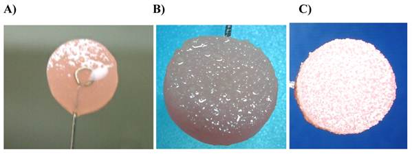
After 24 h, 48 h, and 5 days, the biofilm-coated discs (Fig. 1) were washed with saline to remove unbound bacteria. The discs were then immersed in 4 mL of saline and, to detach the bacteria from the surface, mixed for 2 min with Elektro.mag MIG and sonicated for 1 min using an ultrasonic water bath (Elma, Singen, Germany). Samples from the suspensions were diluted in saline (10-1, 10-2, 10-3, and 10-4) and 0.1 mL was plated on MSBA for the enumeration of S. mutans and S. sobrinus. Bacterial viability was determined using conventional culture methods (14,15,16,17,18). The plates were incubated for 48 h at 37°C under aerobic conditions supplemented with 5% CO2. The number of colony-forming units (CFU/mL) on suitably diluted plates was determined. Each dilution was plated in triplicate.
Biofilms formed on varnish-coated discs after 24 h (A), 48 h (B), and 5 days (C)
Fluoride analysis of the monobiofilms
Newly constructed S. mutans and S. sobrinus monobiofilms were used to determine the fluoride content of the biofilms. First, the wet weights of the biofilms were determined at 24 h, 48 h, and 5 days after carefully removing the deposits with a sterile scalpel, placing them in pre-weighed microcentrifugre tubes, and allowing them to stand for 5 min in air at room temperature. The plaque and microcentrifugre tubes were weighed and the final weights recorded. By subtracting the weight of the microcentrifuge tubes from the final value, the wet weights were obtained as described (19).
The fluoride content in the biofilm samples at different time points was measured according to the microdiffusion method described by Taves (20). The samples (dissolved in 1 mL of deionized water) were placed in 10-cm plastic dishes. Next, 2 mL of 4 M perchloric acid (HClO4) saturated with hexamethyldisiloxane was added to the samples. A trapping solution, 50 µL of 0.5 M sodium hydroxide (NaOH) in a 3-cm plastic dish, was placed in the 10-cm plastic dish and immediately sealed. The samples were placed in a diffusion vessel for 18 h at room temperature with agitation (100 rpm). Next, the 3-cm plastic dishes were dried at 65ºC for 2 h. When the samples reached room temperature, 50 µL of 0.5 M HCl, 400 µL of acetate buffer, and 450 µL of TISAB II were added. The fluoride concentrations, expressed as μg/g, were calculated after measurement with a fluoride-sensitive electrode (96-09; Orion Research, Cambridge, MA, USA), standardized in the range of 0.0625-1 ppm. All determinations were performed in duplicate. The data are presented as µg/g wet sample weight.
RESULTS
The antibacterial effects of the varnishes on the viability of the S. mutans and S. sobrinus monobiofilms are shown in Table 1. A marked increase in the viable counts of S. mutans and S. sobrinus during the test period was observed in all test groups, which was statistically significant. As shown in Table 1, there was a significant relationship between the tested materials at 24 h and 48 h.
Antibacterial effects of the varnishes on S. mutans and S. sobrinus biofilm viability; comparison of viable counts of bacteria between the test groups and between the two biofilms.
| Viable counts of S. mutans and S.sobrinus (106 CFU/mL) | ||||||||
|---|---|---|---|---|---|---|---|---|
| 24 h (S.mutans) | 24 h (S.sobrinus) | 48 h (S.mutans) | 48 h (S.sobrinus) | 5 days (S.mutans) | 5 days (S.sobrinus) | p- value (S.mutans) | p-value (S.sobrinus) | |
| Bifluoride 12 | 1,29±0.52 a,b,c,* | 0.707±0.276 a,b,c, * | 1450±591.38 a,b,c, * | 1.38±0.18 a,b,c, * | 34285.71±7674.94 * | 5225.71±3296.65 * | 0.001 | 0.001 |
| Fluor Protector | 0.003±0.002 a,d,e, # | 0.313±0.184 a,d,e, # | 125.71±62.14 a,d,e, # | 3.55±1.42 a,d,e, # | 31285.71±5186.98 # | 3687.14±1186.71 # | 0.001 | 0.001 |
| Fluor Protector + Cervitec | 0.004±0.002 b,d,f, £ | 0.083±0.029 b,d,f,£ | 204.29±61.33 b,d,f, £ | 1.31±0.13 b,d,f, £ | 32714.29±8731.44 £ | 3937.14±1012.81 £ | 0.001 | 0.001 |
| Control | 2.29±0.52 c,e,f, “ | 1.01±0.23 c,e,f, “ | 2440±401.95 c,e,f, “ | 5.05±1.11 c,e,f, “ | 32571.43±6604.47 ” | 5728.57±1998.93 “ | 0.001 | 0.001 |
| p-value | 0.0001 | 0.0001 | 0.0001 | 0.0001 | 0.921 | 0.161 | ||
p < 0.001 (Kruskal-Wallis and Friedman tests were used), p < 0.05 (Dunn's test).
a Bifluoride 12 / Fluor Protector, p < 0.05 (24 h), p < 0.05 (48 h); b Bifluoride 12 / Fluor Protector + Cervitec, p > 0.05 (24 h), p > 0.05 (48 h); c Bifluoride 12 / Control, p > 0.05 (24 h), p > 0.05 (48 h); d Fluor Protector / Fluor Protector + Cervitec, p > 0.05 (24 h), p > 0.05 (48 h); e Fluor Protector / Control, p < 0.001 (24 h), p < 0.001 (48 h); f Fluor Protector + Cervitec / Control, p < 0.01 (24 h), p < 0.01 (48 h) for S. mutans biofilm.
a Bifluoride 12 / Fluor Protector, p > 0.05 (24 h), p > 0.05 (48 h); b Bifluoride 12 / Fluor Protector + Cervitec, p < 0.01 (24 h), p > 0.05 (48 h); c Bifluoride 12 / Control, p > 0.05 (24 h), p < 0.01 (48 h); d Fluor Protector / Fluor Protector + Cervitec, p > 0.05 (24 h), p < 0.05 (48 h); e Fluor Protector / Control, p < 0.05 (24 h), p > 0.05 (48 h); f Fluor Protector + Cervitec / Control, p < 0.001 (24 h), p < 0.001 (48 h) for S. sobrinus biofilm.
* Bifluoride 12, p = 0.047 (24 h), p = 0.002 (48 h), p = 0.002 (5 days); Fluor Protector, # p = 0.001 (24 h), p = 0.002 (48 h), p = 0.002 (5 days); Fluor Protector + Cervitec, £ p = 0.002 (24 h), p = 0.002 (48 h), p = 0.002 (5 days); Control, “ p = 0.002 (24 h), p = 0.002 (48 h), p = 0.002 (5 days) for comparison of viable bacterial counts between the two biofilms.
Comparisons of viable counts of bacteria between the test groups for biofilms of both species are evaluated by using Dunn's test. Among the test groups, Fluor Protector + Cervitec group had the lowest values of viable counts in both species at 24 and 48 h in comparison to control group. In comparison Fluor Protector group with the control groups the same effect was observed at 24 h in both species. The Fluor Protector group exhibited prolonged antibacterial effect in S.mutans biofilm than S. sobrinus biofilm. A comparison of viable bacterial counts between the two species biofilms is evaluated with Mann-Whitney U-test. All of the dental varnishes tested significantly reduced the viable cell count; however, the effect was stronger for S. sobrinus than for S. mutans.
The fluoride concentrations in both monobiofilms are presented in Table 2. In all groups, the highest amount of fluoride release occurred during the first 24 h, followed by a significant decrease over the next 4 days. As shown in Table 2, there was a significant relationship between the tested materials at all time intervals.
Fluoride concentrations in S. mutans and S. sobrinus biofilms and comparison of fluoride concentrations between the test groups and between the two biofilms.
| 24 h (S.mutans) | 24 h (S.sobrinus) | 48 h (S.mutans) | 48 h (S.sobrinus) | 5 days (S.mutans) | 5 days (S.sobrinus) | p- value (S.mutans) | p-value (S.sobrinus) | |
|---|---|---|---|---|---|---|---|---|
| Bifluoride 12 | 163.39±36.08 a,b,c,* | 152.73±20.4 a,b,c, * | 18.44±2.66 a,b,c, * | 35.3±5.31 a,b,c, * | 7.7±0.97 a,b,c, * | 7.34±1.43 a,b,c, * | 0.001 | 0.001 |
| Fluor Protector | 6.73±1.98 a,d,e, # | 14.01±2.51 a,d,e, # | 3.03±0.7 a,d,e, # | 3.83±0.7 a,d,e, # | 0.89±0.23 a,d,e, # | 0.5±0.2 a,d,e, # | 0.001 | 0.001 |
| Fluor Protector + Cervitec | 10.99±2.19 b,d,f, £ | 6.66±1.13 b,d,f, £ | 2.53±0.65 b,d,f, £ | 3.4±0.6 b,d,f, £ | 0.7±0.26 b,d,f, £ | 0.5±0.12 b,d,f,,£ | 0.001 | 0.001 |
| Control | 1.4±0.41c,e,f, “ | 1.73±0.49 c,e,f, “ | 0.99±0.24 c,e,f, “ | 1.07±0.34 c,e,f, “ | 0.21±0.07 c,e,f, “ | 0.27±0.08 c,e,f, “ | 0.001 | 0.002 |
| p-value | 0.0001 | 0.0001 | 0.0001 | 0.0001 | 0.0001 | 0.0001 |
p < 0.001 (Kruskal-Wallis and Friedman tests were used), p < 0.05 (Dunn's test).
a Bifluoride 12 / Fluor Protector, p < 0.05 (24 h), p > 0.05 (48 h), p > 0.05 (5 days); b Bifluoride 12 / Fluor Protector + Cervitec, p > 0.05 (24 h), p < 0.05 (48 h), p < 0.05 (5 days); c Bifluoride 12 / Control, p < 0.001 (24 h), p < 0.001 (48 h), p < 0.001 (5 days); d Fluor Protector / Fluor Protector + Cervitec, p > 0.05 (24 h), p > 0.05 (48 h), p > 0.05 (5 days); e Fluor Protector / Control, p > 0.05 (24 h), p < 0.05 (48 h), p < 0.05 (5 days); f Fluor Protector + Cervitec / Control, p < 0.05 (24 h), p > 0.05(48 h), p > 0.05 (5 days) for S. mutans biofilm.
a Bifluoride 12 / Fluor Protector, p > 0.05 (24 h), p > 0.05 (48 h), p < 0.05 (5 days); b Bifluoride 12 / Fluor Protector + Cervitec, p < 0.01 (24 h), p < 0.05 (48 h), p > 0.05 (5 days); c Bifluoride 12 / Control, p < 0.001 (24 h), p < 0.001 (48 h), p < 0.001 (5 days); d Fluor Protector / Fluor Protector + Cervitec, p > 0.05 (24 h), p > 0.05 (48 h), p > 0.05 (5 days); e Fluor Protector / Control, p < 0.01 (24 h), p < 0.05 (48 h), p > 0.05 (5 days); f Fluor Protector + Cervitec / Control, p > 0.05 (24 h), p > 0.05(48 h), p > 0.05 (5 days) for S. sobrinus biofilm.
Bifluoride 12, * p = 0.848 (24 h), p = 0.002 (48 h), p = 0.749 (5 days); Fluor Protector, # p = 0.002 (24 h), p = 0.047 (48 h), p = 0.014 (5 days); Fluor Protector + Cervitec, £ p = 0.003 (24 h), p = 0.034 (48 h), p = 0.078 (5 days); Control, “ p = 0.178 (24 h), p = 0.653 (48 h), p= 0.184 (5 days) for comparison of fluoride concentrations between the two biofilms.
A comparison of fluoride concentrations between the test groups in both biofilms is evaluated by using Dunn's test. Among the test groups, Bifluoride 12 showed the highest fluoride concentration in both biofilms in comparison to control groups during the test period. A statistically significant difference was found only at 24 h S.mutans biofilm at Fluor Protector + Cervitec group in comparison to control group. Fluor Protector group showed statistically significant difference in comparison to control group at 48 h and 5 days in S.mutans biofilm, meanwhile same statistical relation was observed at 24 h and 48 h in S.sobrinus biofilm.
A comparison of fluoride concentrations between the two biofilms is evaluated with Mann-Whitney U-test. The fluoride concentrations in the S. sobrinus biofilms exposed to Bifluoride 12 were significantly higher than in the S. mutans biofilms at 48 h. The fluoride concentrations in the S. sobrinus biofilms in the Fluor Protector group were significantly higher than in the S. mutans biofilms at 24 and 48 h. After 5 days, the fluoride concentration in the S. mutans biofilms in the Fluor Protector group was significantly higher than in the S. sobrinus biofilms. In the Fluor Protector + Cervitec group, the fluoride concentration in the S. mutans biofilms at 24 h was significantly higher than that in the S. sobrinus biofilms; however, at 48 h, the fluoride concentration in the S. sobrinus biofilms was higher than that in the S. mutans biofilms.
A comparison of the viable counts of bacteria and fluoride concentrations in both biofilms is presented in Table 3. A negative correlation was detected between the fluoride concentration and antibacterial effect in all study groups with both biofilms. Over time, as the fluoride concentration decreased, the viability of S. mutans and S. sobrinus in the biofilms increased.
Comparison of viable counts of bacteria and fluoride concentrations in both biofilms over time (24 h to 5 days)
| Fluoride concentration in monobiofilm (µg/g) | S. mutans | S. sobrinus | |
|---|---|---|---|
| Viable counts of bacteria (CFU/mL) | Viable counts of bacteria (CFU/mL) | ||
| Bifluoride 12 | r | -0.933 | -0.685 |
| p | 0.0001 | 0.001 | |
| N | 21 | 21 | |
| Fluor Protector | r | -0.911 | -0.826 |
| p | 0.0001 | 0.0001 | |
| N | 21 | 21 | |
| Fluor Protector + Cervitec | r | -0.96 | -0.91 |
| p | 0.0001 | 0.0001 | |
| N | 21 | 21 | |
| Control | r | -0.793 | -0.845 |
| p | 0.0001 | 0.0001 | |
| N | 21 | 21 | |
(Spearman's correlation test)
Statistical analysis
The data were analyzed using GraphPad Prism software (ver. 3). A statistical analysis of the antibacterial effect of the different varnishes on S. mutans and S. sobrinus biofilms was performed by first subjecting the CFU numbers to a logarithmic transformation. The numbers of colonies are presented as millions. The Kruskal-Wallis test was used to compare the experimental groups with both monobiofilms. The Friedman test was used to compare the experimental groups over time. Dunn's test was then used to compare the groups according to time interval. The Mann-Whitney U-test was used to compare the monobiofilms with each other and Spearman's correlation analysis was used to compare the viability of the colonies and fluoride concentration in the monobiofilms. The mean and standard deviations of the fluoride concentrations in both biofilms are presented. The level of significance was set at p < 0.05.
DISCUSSION
Antimicrobial agents are available in different formulations, including toothpastes, mouthwashes, sprays, and gels. More recently, antimicrobials have been incorporated into a variety of sustained-release systems, including varnishes. The rationale is simply to deliver the active agent at the tooth surface for prolonged periods of time. The impact of antiseptic varnishes on the microbiota, in particular on cariogenic bacteria, has been well-documented through clinical trials and in vitro studies (18,21,22). One of the main pharmaceutical goals in preventing dental caries is to decrease the viable biofilm mass.
The present study was designed to gain information on biofilm formation with varnishes containing Fluor Protector, Bifluoride 12, and a combination of Fluor Protector + Cervitec (1:1). To our knowledge, there is no previous published report of biofilm formation on varnishes containing different levels of fluoride and chlorhexidine in both S. mutans and S. sobrinus biofilms measured at different time points. The effects of antimicrobials on oral biofilms in vitro, in situ, and in vivo can only be compared if the time between last treatment and sampling is taken into consideration (18).
In our study, when the antibacterial effects of the test materials were evaluated, of the three varnishes tested, Fluor Protector + Cervitec had the highest inhibitory effect against S. mutans and S. sobrinus biofilms, while Bifluoride 12 had the lowest inhibitory effect during the experimental period, although it had the highest fluoride concentration.
These differences may be explained by the characteristics of the different varnishes and mechanisms of action. It is well-established that chlorhexidine has antimicrobial activity against most bacterial species found in the oral cavity. Chlorhexidine is a bis-biguanide with antibacterial, anticariogenic, and remineralizing actions and few toxic effects. Chlorhexidine acts by damaging the cell membrane of prokaryotes and disrupting their cytoplasmic constituents. Cell death occurs due to the rapid accumulation of metal ions inside the cells as they become more permeable. Several clinical studies have reported that chlorhexidine-containing varnishes produce long-lasting (up to several months) suppression of S. mutans (10,11). Sustained-release systems, including varnishes, generally show an initial burst, with rapid release of the active agent, followed by a slower phase of release (7,18). In our experiments, the activity decreased over the experimental period.
Bifluoride 12 has a higher viscosity than the other test materials, which may have resulted in a thicker layer on the acrylic surface. The adherence of the bacteria to this surface may have been easier than that in the other groups.
The statistically significant difference between the viable counts of bacteria in the Bifluoride 12 group after 24 h and 5 days shows that this varnish affected bacterial viability at 24 h but that this effect was minor when compared with the other test groups.
Fluor Protector contains the polyurethane-based compound difluorosilane, has a low pH, and formed a thin transparent film on the disc surface. Although Fluor Protector contained a lower fluoride concentration than Bifluoride 12, its antibacterial effect was better, and this may be explained by the silane content. The addition of Cervitec to Fluor Protector increased the antibacterial effect and efficacious time of Fluor Protector against both biofilms.
When the effects of dental varnishes on dental biofilms were examined, the thickness of the biofilm increased between 24 h and 5 days. The fluoride concentration peaked after 24 h then decreased while the thickness of the biofilm increased.
In this study, the wet weights of the monobiofilms of S. mutans and S. sobrinus increased on the discs with dental varnishes over 5 days. The bacteria that survived and continued to grow produced an extracellular matrix. It is thought that, as the biofilm thickness was increasing during the 5 days, the penetration of antimicrobials through the biofilm could be blocked and that pH differences in the plaque layers could cause a decrease in the antibacterial efficacy of the test varnishes.
When the relationships between the fluoride concentrations and antibacterial effects were examined in the study groups, it was found that as the fluoride concentration decreased, the viable bacterial counts increased. Thus, it is possible that the rapid release of fluoride from the varnishes resulted in remaining concentrations that may have been too low to exert an antibacterial effect or to inhibit biofilm formation. Similar results were observed with both biofilms. In this study, the fluoride concentrations in the Bifluoride 12 group in both biofilms were significantly higher at 24 h; this result was considered a “burst effect.”
Although the highest fluoride concentrations were found in both biofilms with Bifluoride 12, the highest viable counts of bacteria were also observed in these films. This result requires some discussion of the antibacterial effects of fluoride. However, it must be emphasized that this finding does not exclude the possibility of an inhibitory effect of fluoride varnishes on the rate of acid production in biofilms. Fluoride may interfere with bacterial metabolism and inhibit bacterial growth (8,9). The results of this study support the limited antibacterial effect of fluoride. The antibacterial effect within the study groups could be explained by the antibacterial agents in the dental varnishes. In discussing the results of this study, the experimental methods, environmental pH, and pH of the test varnishes are parameters that should be considered.
Our experimental model mimics several of the environmental conditions in the oral cavity such as saliva, bacteria and in situ polysaccharide production which affect bacterial adhesion to surfaces. Possible limitation of this study may include using monospecific biofilms of S. mutans and S. sobrinus. Monospecific biofilms of late-colonizing streptococci or mixed culture biofilms should be used to confirm these results.
Conclusions
In conclusion, in vitro oral biofilm models represent a valuable tool for studying and testing chemical agents. The present results indicate that the Fluor Protector + Cervitec group exhibited the greatest antibacterial effect, which is important in delaying bacterial colonization and biofilm development. While Bifluoride 12 showed the smallest inhibitory effect, it had the highest fluoride concentration. For all groups, the highest amount of fluoride release was observed during the first 24 h, and was followed by a significant decrease over the following 4 days; as the fluoride concentration decreased, the viable counts of the bacteria increased. Further investigation should be carried out to confirm these results and to develop strategies for using such products to prevent dental caries.
Acknowledgements
This study was supported by The Research Support Unit of Istanbul University, project no. T-535/21102004.
Conflict of Interest
The authors have declared that no conflict of interest exists.
References
1. Fejerskov O, Kidd EAM. Clinical cariology and operative dentistry in the twenty-first century. In: (ed.) Fejerskov O, Kidd E. Dental Caries; The Disease and Its Clinical Management. Denmark, Copenhagen: Blackwell Publishing Ltd. 2003:179-188
2. Nisengard RJ, Newman MG. Oral Microbiology and Immunology, second edition. Philadelphia: WB Saunders Company. 1994
3. Schachtele CF. Dental Caries: Prevention and control. Stallard RE. A Textbook of Preventive Dentistry, 2nd ed. Philadelphia: WB Saunders Company. 1982:241-54
4. Shaw JH. Etiology of dental caries: Prevention and control. Stallard RE. A Textbook of Preventive Dentistry, 2nd ed. Philadelphia: WB Saunders Company. 1982:32-49
5. Bowden GHW, Li YH. Nutritional influences on biofilm development. Adv Dent Res. 1997;11:81-99
6. Scheie AA. Modes of currently known chemical antiplaque agents other than chlorhexidine. J Dent Res. 1989;68(Suppl):S1609-1616
7. Scheie AA. The role of antimicrobials. In: (ed.) Fejerskov O, Kidd E. Dental Caries: The Disease and Its Clinical Management. Denmark, Copenhagen: Blackwell Publishing Ltd. 2003:179-188
8. Bradshaw DJ, Marsh PD, Hodgson RJ, Visser JM. Effect of glucose and fluoride on competition and metabolism within in vitro dental bacterial communities and biofilms. Caries Res. 2002;36:81-86
9. Horowitz HS, Ismail AI. Topical fluorides in caries prevention. Fejerskov O, Ekstrand J, Burt BA. Fluoride In Dentistry, 2nd ed. Copenhagen: Munksgaard. 1996:311-327
10. Zaura-Arite E, Ten Cate JM. Effects of fluoride and chlorhexidine containing varnishes on plaque composition and on demineralization of dentinal grooves in situ. Eur J Oral Sci. 2000;108:154-161
11. Sorvari R, Happonen SS, Luoma H. Efficacy of chlorhexidine solution with fluoride varnishing in preventing enamel softening by Streptococcus mutans in an artificial mouth. Scand J Dent Res. 1994;102:206-209
12. Gold OG, Jordan HV, Van Houte J. A selective medium for Streptococcus mutans. Arch Oral Biol. 1973;18:1357-1364
13. Steinberg D, Beeman D, Bowen WH. The effect of delmopinol on glucosyltransferase adsorbed on to a saliva-coated hydroxyapatite. Arch Oral Biol. 1992;37:33-38
14. Guggenheim B, Giertsen E, Schupbach P, Shapiro S. Validation of an in vitro biofilm model of supragingival plaque. J Dent Res. 2001;80:363-370
15. Fraud S, Maillard JY, Kaminski MA, Hanlon GW. Activity of amine oxide against biofilms of Streptococcus mutans: a potential biocide for oral care formulations. J Antimicrob Chemother. 2005;56:672-677
16. Hardie JM. Oral microbiology: current concepts in the microbiology of dental caries and periodontal disease. Br Dent J. 1992;172:271-278
17. Hope CK, Wilson M. Comparison of the interproximal plaque removal efficacy of two powered toothbrushes using in vitro oral biofilms. Am J Dent. 2002;15:7-11
18. Steinberg D, Rozen R, Klausner EA, Zachs B, Friedman M. Formulation, development and characterization of sustained release varnishes containing amine and stannous fluorides. Caries Res. 2002;36:411-416
19. Rajtboriraks D, Nakornchai S, Bunditsing P, Surarit R, Iemjarern P. Plaque and saliva fluoride levels after placement on fluoride releasing pit and fissure sealants. Paediatric Dentistry. 2004;26:63-66
20. Taves DR. Separation of fluoride by rapid diffusion using hexamethyldisiloxane. Talanta. 1968;15:969-974
21. Munshi AK, Reddy NN, Shetty V. A comparative evaluation of three fluoride varnishes: an in-vivo study. J Indian Soc Pedo Prev Dent. 2001;19:92-102
22. Shapiro S, Giertsen E, Guggenheim B. An in vitro oral biofilm model for comparing the efficacy of antimicrobial mouth rinses. Caries Res. 2002;36:93-100
Author contact
![]() Corresponding author: Dr. Arzu Pinar Erdem, Department of Pedodontics, Faculty of Dentistry, Istanbul University, 34093 Capa/Istanbul/Turkey. Tel: +90212 4142020/30317,Telefax: +90212 5310515, e-mail: pinararzuerdemcom.
Corresponding author: Dr. Arzu Pinar Erdem, Department of Pedodontics, Faculty of Dentistry, Istanbul University, 34093 Capa/Istanbul/Turkey. Tel: +90212 4142020/30317,Telefax: +90212 5310515, e-mail: pinararzuerdemcom.