Impact Factor ISSN: 1449-1907

 Global reach, higher impact
Int J Med Sci 2022; 19(6):1047-1048. doi:10.7150/ijms.73905 This issue Cite
Erratum
Co-culture with Endothelial Progenitor Cells promotes the Osteogenesis of Bone Mesenchymal Stem Cells via the VEGF-YAP axis in high-glucose environments: Erratum
Peilian Wu1,2,3*, Xia Zhang1,4*, Yun Hu1,2,3, Dongrong Liu1,2,3, Jinlin Song1,2,3, Wenjie Xu1,2,3, Hao Tan1,2,3, Rui Lu1,2,3, Leilei Zheng1,2,3 ![]()
1. The Affiliated Stomatology Hospital, Chongqing Medical University, Chongqing, 401147, China.
2. Chongqing Key Laboratory of Oral Diseases and Biomedical Sciences, Chongqing, 401147, China.
3. Chongqing Municipal Key Laboratory of Oral Biomedical Engineering of Higher Education, Chongqing, 401147, China.
4. West china dental hospital of Chongqing, Chongqing, 401147, China.
*These authors contributed equally to this work.
Published 2022-6-12
Corrected-article in Int J Med Sci, Volume 18, 1628
In our paper, Figure 1B-b and Figure 1 should be corrected as follows.
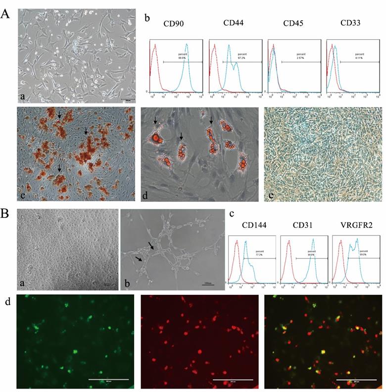
Corrected Figure 1B-b.
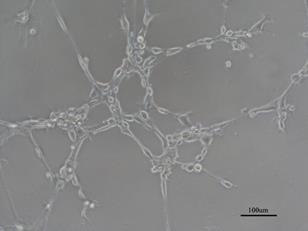
Corrected Figure 1. Characterization of BMSCs and EPCs. A) Isolation and differentiate of BMSCs a morphology of BNSCs,P0×100; b) Cell surface markers of BMSCs c) Alizarin red staining of BMSCs after osteogenic induction (×100; Black arrows indicate calcium deposits); d) Oil red O staining of BMSCs after adipogenic induction (×200; The black arrow indicates lipid droplets); e Alcain staining of BMSCs after chrondrogenic induction (×100). B) Isolation and identification EPCs a) morphology of epc, P1×100; b) Matrigel tubule formation experiment of EPCs (×100.Black arrow indicates tubule). c) Cell surface markers of EPCs d) Double fluorescence staining experiment of EPCs, (×200.green: FITC-UEA-1 red: DiI-Ac-LDL, yellow: fluorescence coincidence).
Author contact
![]() Corresponding author: Leilei Zheng, DDS, Ph.D. The Affiliated Stomatology Hospital, Chongqing Medical University. No.426 Songshibei Road,Yubei District, Chongqing, 401147. E-mail: zhengleileicqmucqmu.edu.cn.
Corresponding author: Leilei Zheng, DDS, Ph.D. The Affiliated Stomatology Hospital, Chongqing Medical University. No.426 Songshibei Road,Yubei District, Chongqing, 401147. E-mail: zhengleileicqmucqmu.edu.cn.